Pleural Mesothelial Cells, Https Onlinelibrary Wiley Com Doi Pdf 10 1002 Dc 20938
Pleural mesothelial cells Indeed lately is being hunted by consumers around us, perhaps one of you. People now are accustomed to using the internet in gadgets to view video and image data for inspiration, and according to the title of this article I will talk about about Pleural Mesothelial Cells.
- Lrrn4 And Upk3b Are Markers Of Primary Mesothelial Cells
- Pleural Effusion Flashcards Cram Com
- Mesothelial Cell In Pleural Fluid Stock Photo Picture And Royalty Free Image Image 65541205
- Buy Effects Of Anti Dna Antibodies On Pleural Mesothelial Cells In Vitro Studies To Explore The Pathogenetic Mechanism Of Pulmonary Lupus Book Online At Low Prices In India Effects Of Anti Dna Antibodies
- Atypical Mesothelial Cells In Pleural Effusion Youtube
- Mesothelial Cell In Pleural Fluid Stock Photo Picture And Royalty Free Image Image 65541216
Find, Read, And Discover Pleural Mesothelial Cells, Such Us:
- Effusions Abdominal Thoracic And Pericardial Veterian Key
- Surgical Pathology Of Non Neoplastic Conditions Of The Pleura Pericardium And Peritoneum Chapter 6 Practical Pathology Of Serous Membranes
- Figure 1 From Pathophysiology Of The Pleura Semantic Scholar
- Mesothelial Cell In Pleural Fluid Stock Photo Picture And Royalty Free Image Image 65541216
- Top Pdf Mesothelial Cells 1library
- Coloring Sheets For Girls Easy
- Liam Tribe Simmons
- Iowa Mesothelioma Prognosis
- Family Of Lawyers
- Fictional Law Firm Names
If you are looking for Fictional Law Firm Names you've come to the perfect place. We have 104 images about fictional law firm names including pictures, pictures, photos, wallpapers, and much more. In these page, we also have number of images available. Such as png, jpg, animated gifs, pic art, logo, black and white, translucent, etc.

Non Neoplastic Reactive Mesothelial Cells Dog With Non Neoplastic Download Scientific Diagram Fictional Law Firm Names
There are certain cells that line the pleura the thin double layered lining which covers the lungs chest wall and diaphragm which are known as mesothelial cellsother than the pleura mesothelial cells also form a lining around the heart pericardium and the internal surface of the abdomen peritoneum.

Fictional law firm names. Pleural mesothelial cells pmcs undergo a process called mesothelial mesenchymal transition mesomt by which pmcs acquire a profibrotic phenotype characterized by cellular enlargement and elongation increased expression of a smooth muscle actin a sma and matrix proteins including collagen 1. The pleural mesothelial cell pmc is the most common cell in the pleural space and is the primary cell that initiates responses to noxious stimuli. Transformed normal pleural mesothelial cells met5a or pmcs from patients with ipf were seeded at a density of 1 10 6 cellsml.
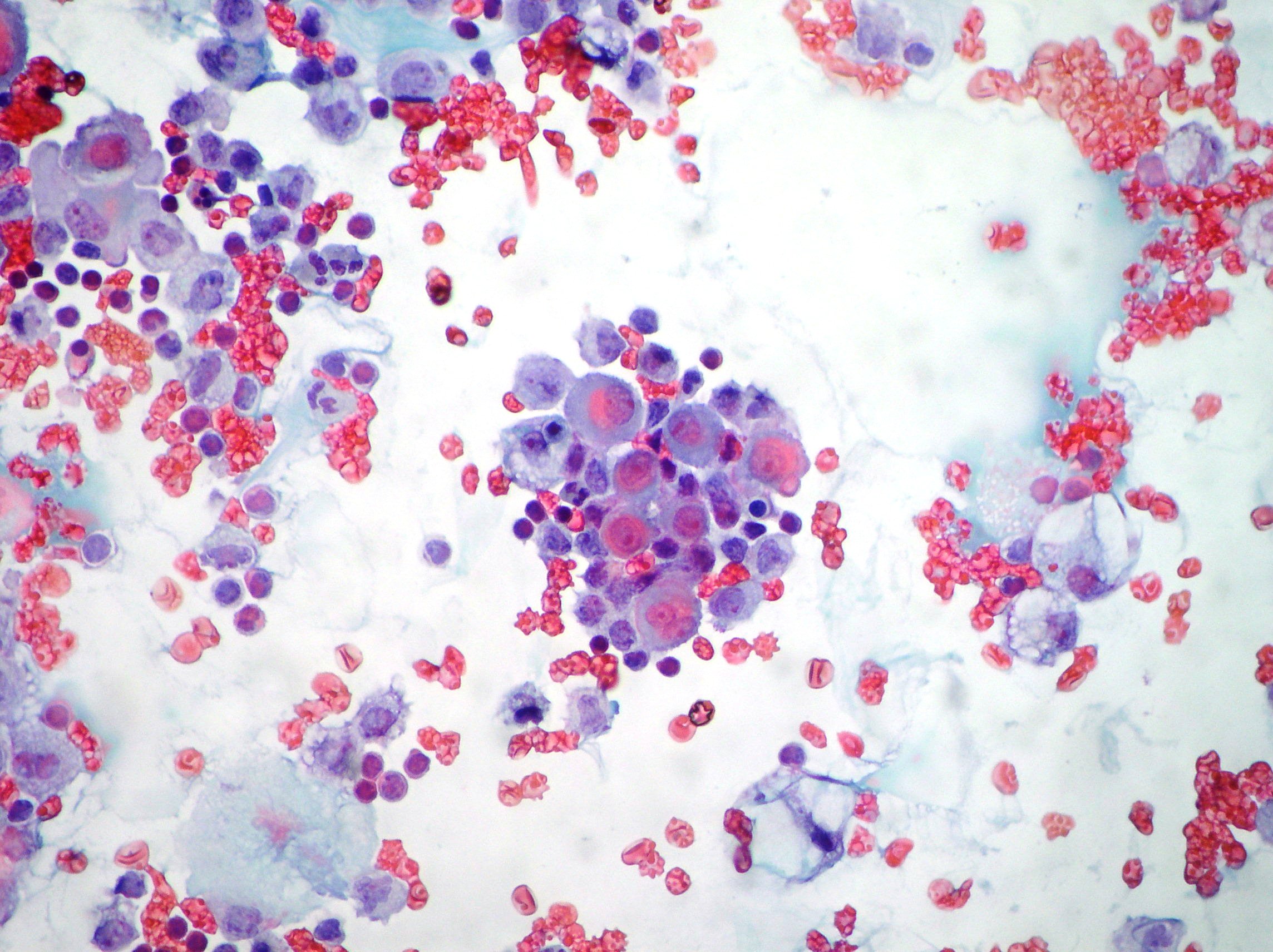
Numerous mesothelial cells are seen in this pleural fluid from a dog with a transudative effusion with concurrent diapedesis of red blood cells or hemorrhage. The mesothelial cells have central round nuclei with a moderate amount of light purple cytoplasm and a corona or fringe to the cytoplasmic borders. Pmcs are metabolically active cells that maintain a dynamic state of homeostasis in the pleural space.
Mesothelial cells in pleural fluid. Sigma aldrich in a 31 proportion and seeded in individual wells in a 24 well culture dish. Although mesomt contributes to pleural.
Mesothelial cell is the predominant cell type in the pleural cavity see mesothelial cells. There are many possible causes of pleural effusions. Mesothelial cells are found in variable numbers in most effusions but their presence at greater than 5 of total nucleated cells makes a diagnosis of tb less likely.
No significant differences have been identified between the mesothelial cells in the visceral or parietal pleura or among those in the pleural peritoneal or pericardial cavities. Pleural effusions are a build up of fluid in the cavity between the two layers of the pleura the pleural mesothelium and is influenced by substances secreted by pleural mesothelial cells. Markedly increased numbers of.
The lung can get stuck in the chest cavity or become trapped if the pleural lining is impaired during healing.

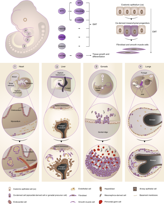
Mesothelial To Mesenchyme Transition As A Major Developmental And Pathological Player In Trunk Organs And Their Cavities Communications Biology Fictional Law Firm Names
More From Fictional Law Firm Names
- Mesothelioma Prognosis Stages
- Is 2 Asbestos Dangerous
- Zombie Coloring Pages Halloween
- Malignant Pleural Effusion End Of Life
- Unicorn Pusheen Coloring Pages
Incoming Search Terms:
- Mesothelioma In A Pleural Fluid Specimen Atypical Mesothelial Cells Download Scientific Diagram Unicorn Pusheen Coloring Pages,
- Up Regulation Of Ddx39 In Human Malignant Pleural Mesothelioma Cell Lines Compared To Normal Pleural Mesothelial Cells Unicorn Pusheen Coloring Pages,
- Effusions Cytopathology Cellnetpathology Unicorn Pusheen Coloring Pages,
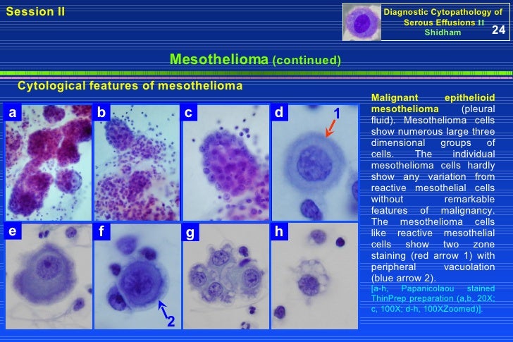
- Mesothelioma Vs Reactive Mesothelial Cells Cytology Creative Art Unicorn Pusheen Coloring Pages,
- The Value Of Cytology And Pleural Biopsy In The Differential Diagnostic Of Nonspecific Pleural Effusions Unicorn Pusheen Coloring Pages,
- Pathology Outlines Mesothelial Unicorn Pusheen Coloring Pages,


