Mesothelial Cells In Pleural Fluid Pictures, Benign Mesothelial Cells In Pleural Fluid Medical Laboratory Hematology Mad Scientist
Mesothelial cells in pleural fluid pictures Indeed lately is being sought by consumers around us, perhaps one of you. People are now accustomed to using the net in gadgets to view video and image information for inspiration, and according to the title of this post I will talk about about Mesothelial Cells In Pleural Fluid Pictures.
- Mesothelial Cell In Pleural Fluid Stock Photo Picture And Royalty Free Image Image 65541204
- Mesothelium Wikipedia
- Pleural Fluid Mast Cells 2
- Unsuspected Multiples Myeloma Presenting As Bilateral Pleural Effusion A Cytological Diagnosis Abstract Europe Pmc
- Mesothelial Cell Pleural Fluid Stock Photo Edit Now 652971211
- Figure 3 From Telomere Repeat Amplification Protocol Trap In Situ Reveals Telomerase Activity In Three Cell Types In Effusions Malignant Cells Proliferative Mesothelial Cells And Lymphocytes Semantic Scholar
Find, Read, And Discover Mesothelial Cells In Pleural Fluid Pictures, Such Us:
- Effusions Mspca Angell
- Benign Pleural Effusion With Ae1 Ae3 Positive Mesothelial Cells Mycytopathology
- A Panel Of Markers For Identification Of Malignant And Non Malignant Cells In Culture From Effusions
- Http Www Api Pt Com Reference Commentary 2015ascope Pdf
- Http Labmed Oxfordjournals Org Content Labmed 29 1 26 Full Pdf
- David Tobing Law Firm
- Mesothelioma Treatment Plan
- Tyrannosaurus Coloring Page
- Mesothelioma Cell Lines
- Dc Law Logo
If you re looking for Dc Law Logo you've arrived at the right location. We ve got 104 images about dc law logo adding images, photos, pictures, backgrounds, and more. In these web page, we also provide variety of images available. Such as png, jpg, animated gifs, pic art, symbol, black and white, transparent, etc.
The main purpose of these cells is to produce a lubricating fluid that is released between layers providing a slippery non adhesive and protective surface to facilitate intracoelomic movement.
Dc law logo. Atypical mesothelial cell proliferation. There are many possible causes of pleural effusions. Pleural fluid stock photos and images 319 matches.
Of 31 exudative effusions with a lymphocytic predominance 30 were due either to tuberculosis or neoplasm. Plasma cell 250 226 mesothelial cell 214 132 lymphoma cell 36 11 this pleural fluid was obtained from a 60 year old male with newly diagnosed carcinoma of the left lung. Find mesothelial cell pleural fluid stock images in hd and millions of other royalty free stock photos illustrations and vectors in the shutterstock collection.
Note the large size and cytoplasmic basophilia of these cells in compari. Common cells present in pleural fluid include neutrophils lymphocytes monocytes mesothelial cells and red blood. Pleural fluid cytological studies showed malignant cells in 33 of 43 patients with effusions due to tumor.
The arrowed cells all represent atypical lymphocytes. Numerous mesothelial cells are seen in this pleural fluid from a dog with a transudative effusion with concurrent diapedesis of red blood cells or hemorrhage. Pleural effusions are a build up of fluid in the cavity between the two layers of the pleura the pleural mesothelium and is influenced by substances secreted by pleural mesothelial cells.
Picture of mesothelial cell in pleural fluid stock photo images and stock photography. Wbc 7400ul and rbc 4000ul. No tuberculous effusions had more than 1 mesothelial cells while most other effusions contained at least 5 mesothelial cells.
Epithelial or lining cells most commonly mesothelial cells1 the appearance and presentation of nucleated cells found in pleural fluid and whether they are considered commonbenign or abnormal is discussed below. Larger clusters of hyperplastic mesothelial cells showing mildly nuclear atypia with small nucleoli. Thousands of new high quality pictures added every day.
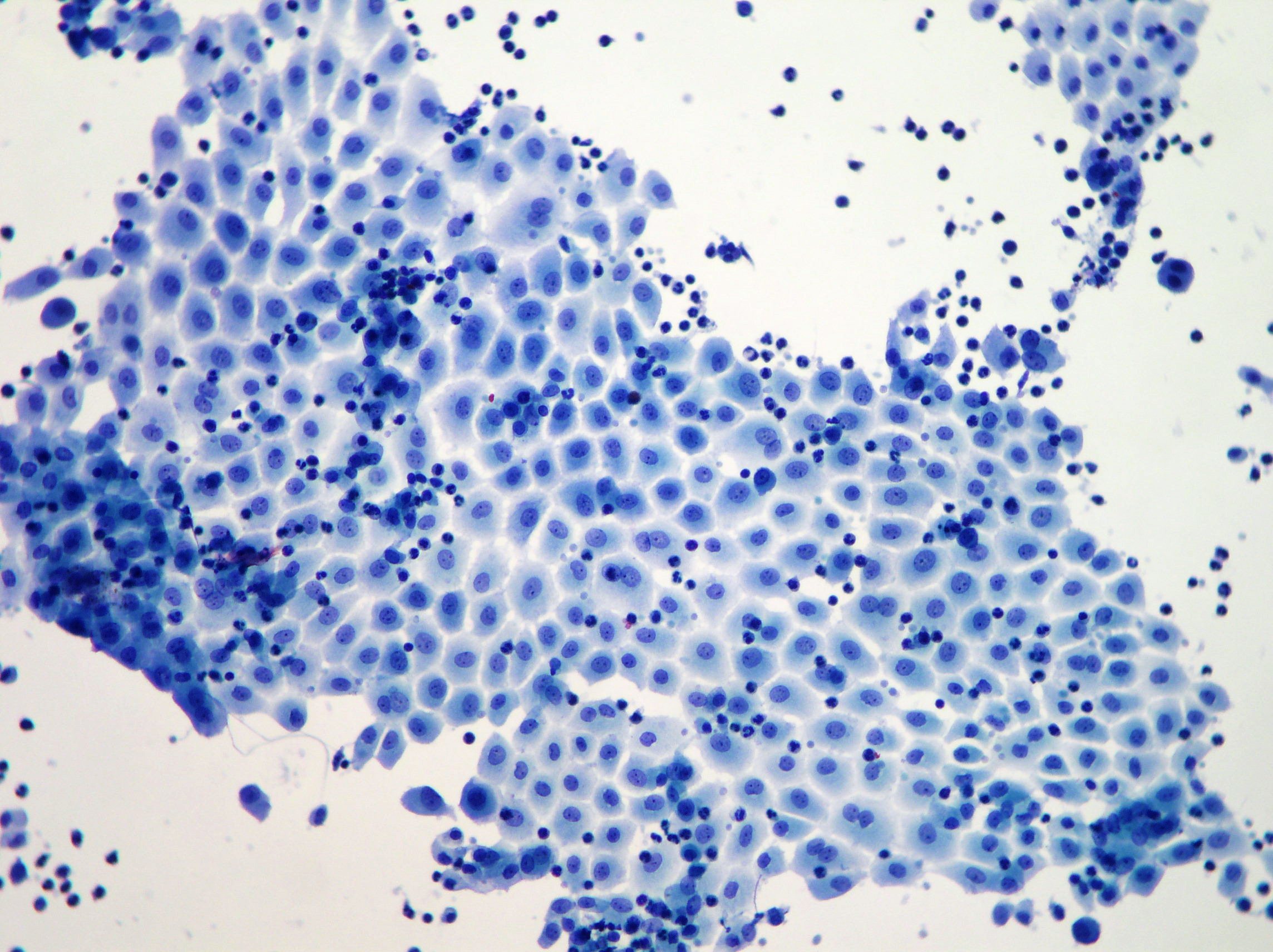
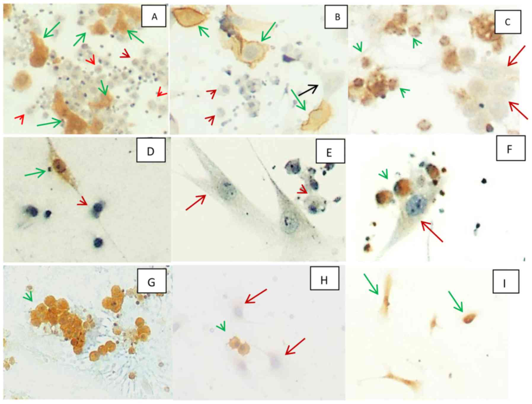
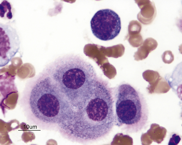
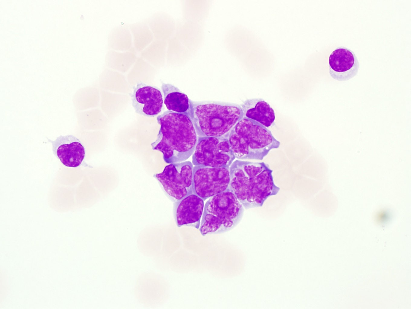
The mesothelial cells have central round nuclei with a moderate amount of light purple cytoplasm and a corona or fringe to the cytoplasmic borders. Hyperplastic mesothelial cells with slightly enlarged nuclei micronucleoli and a clear space or window between adjacent cells present singly and in small clusters.
More From Dc Law Logo
- White Chicks Costume
- Scary Clown Faces Images
- Mesothelioma Mental Health
- How Long Do You Live With Stage 4 Mesothelioma
- Immigration Attorney Myra Jolie
Incoming Search Terms:
- The Value Of Cytology And Pleural Biopsy In The Differential Diagnostic Of Nonspecific Pleural Effusions Immigration Attorney Myra Jolie,
- Cpd From Cloudy To Clear Effusions Made Easy Vet360 Immigration Attorney Myra Jolie,
- Reactive Mesothelial Cells In Pleural Effusion Showing Variation In Download Scientific Diagram Immigration Attorney Myra Jolie,
- Case N 35 Pleural Effusion Mesothelioma Cytology Blog Site Immigration Attorney Myra Jolie,
- Effusions Cytopathology Cellnetpathology Immigration Attorney Myra Jolie,
- Home Immigration Attorney Myra Jolie,


