Pleural Fluid Reactive Mesothelial Cells, Https Encrypted Tbn0 Gstatic Com Images Q Tbn 3aand9gcqipxph4wstcqoym53xfwxvcfrypiwahvqwk3iphwopz5jov0cm Usqp Cau
Pleural fluid reactive mesothelial cells Indeed lately has been hunted by consumers around us, perhaps one of you. People are now accustomed to using the net in gadgets to see video and image data for inspiration, and according to the name of this article I will discuss about Pleural Fluid Reactive Mesothelial Cells.
- Pleural Fluid For Cytology Dr Sachin Kale
- Https Encrypted Tbn0 Gstatic Com Images Q Tbn 3aand9gcqipxph4wstcqoym53xfwxvcfrypiwahvqwk3iphwopz5jov0cm Usqp Cau
- Frontiers Free Floating Mesothelial Cells In Pleural Fluid After Lung Surgery Medicine
- Mesothelioma Vs Adenocarcinoma Cytology Creative Art
- Pleural Fluid Mast Cells 2
- Effusions Cytopathology Cellnetpathology
Find, Read, And Discover Pleural Fluid Reactive Mesothelial Cells, Such Us:
- Benign Effusions Springerlink
- Reactive Mesothelial Cells In Pleural Effusion Showing Variation In Download Scientific Diagram
- Http Labmed Oxfordjournals Org Content Labmed 29 1 26 Full Pdf
- Hjcam Iatrikh Zwwn Syntrofias Hellenic Journal Of Companion Animal Medicine Volume 6 Issue 1 2017 Pleural Effusion In The Cat A Focus On Laboratory Diagnosis
- Effusion Cytology Diagnosis
- Find A Lawyer Nba
- Cubs Pumpkin Carving Templates
- Swear Word Coloring Pages Free Download
- Yoga Poses Yoga Coloring Pages For Kids
- Free Pony Coloring Pages
If you re searching for Free Pony Coloring Pages you've reached the perfect location. We ve got 104 images about free pony coloring pages including images, photos, pictures, backgrounds, and much more. In such page, we additionally provide number of images available. Such as png, jpg, animated gifs, pic art, symbol, blackandwhite, transparent, etc.
Use of pleural fluid n.
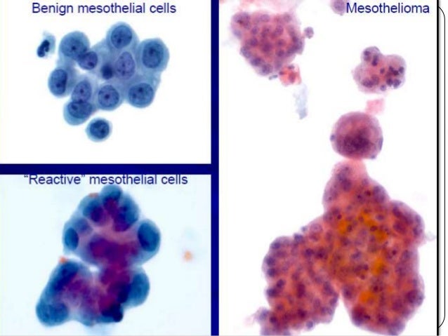
Free pony coloring pages. Some multilayering of parietal mesothelium was occasionally seen in chronic pleurisy and around metastases. There are certain cells that line the pleura the thin double layered lining which covers the lungs chest wall and diaphragm which are known as mesothelial cellsother than the pleura mesothelial cells also form a lining around the heart pericardium and the internal surface of the abdomen peritoneum. During development the mesoderm maintains a complex relationship with the developing endoderm giving rise to the mature lung.
This condition can be due to the presence of a bacterial viral or fungal infection. Mesothelial cells are found in variable numbers in most effusions but their presence at greater than 5 of total nucleated cells makes a diagnosis of tb less likely. Junko ueda takako iwata midori ono mutsuo takahashi comparison of three cytologic preparation methods and immunocytochemistries to distinguish adenocarcinoma cells from reactive mesothelial cells in serous effusion diagnostic cytopathology 101002dc20391 34 1 6 10 2005.
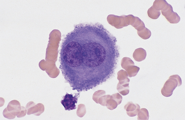
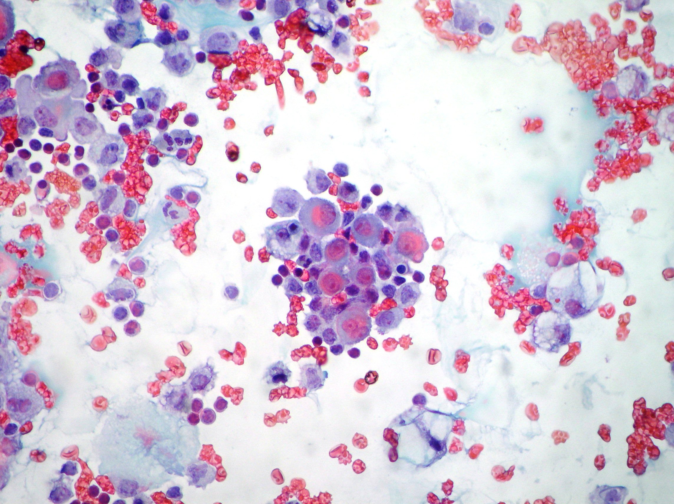
The pleural mesothelium differentiates to give rise to the endothelium and smooth muscle cells via epithelial to mesenchymal transition emt. Mesothelial cells in ascitic fluid mesothelial cells in ascitic fluid the associated tumor antigen 90k is known to possess properties similar. The mesothelial cells have central round nuclei with a moderate amount of light purple cytoplasm and a corona or fringe to the cytoplasmic borders.
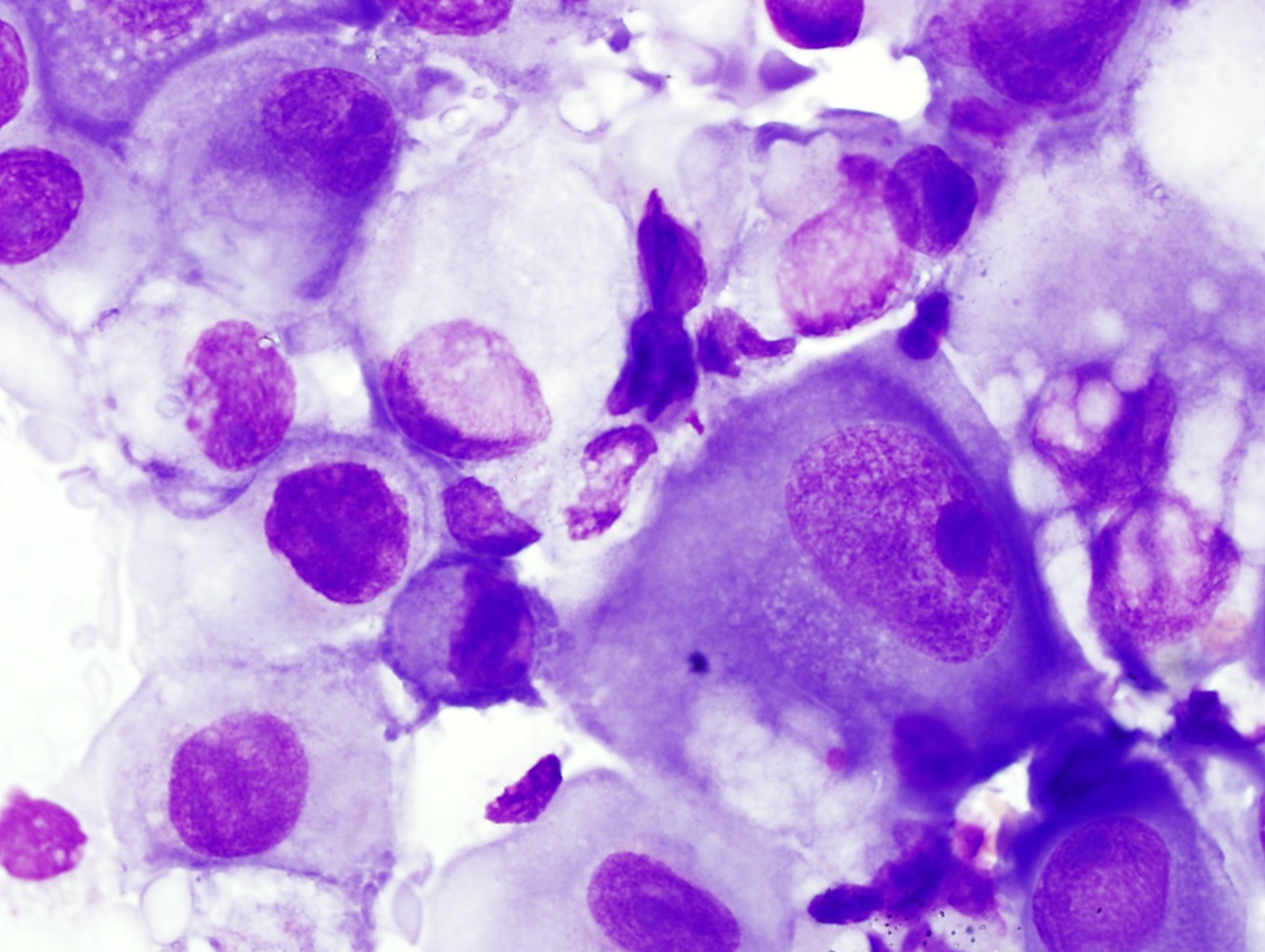
Numerous mesothelial cells are seen in this pleural fluid from a dog with a transudative effusion with concurrent diapedesis of red blood cells or hemorrhage. It can also be the result of trauma or the presence of metastatic tumor. Densely packed mesothelial cells within the pleural space are frequent in benign reactions but densely packed mesothelial cells within the stroma favor a diagnosis of malignancy.
Epithelial or lining cells most commonly mesothelial cells1 the appearance and presentation of nucleated cells found in pleural fluid and whether they are considered commonbenign or abnormal is discussed below. Pleural mesothelial cells pmcs derived from the mesoderm play a key role during the development of the lung. The suggestion that the presence of numerous often very reactive mesothelial cells in pleural aspirate makes the diagnosis of tuberculosis is unlikely confirmed mesothelial cells in pleural fluid.
Reactive mesothelial cells can be found when there is an infection or an inflammatory response present in a body cavity.
More From Free Pony Coloring Pages
- Grumpy Cat Coloring Book
- Oj Lawyer Kardashian
- Riverbank Rink
- Corporate And Commercial Law
- Jacoby Restaurant
Incoming Search Terms:
- Https Encrypted Tbn0 Gstatic Com Images Q Tbn 3aand9gcqipxph4wstcqoym53xfwxvcfrypiwahvqwk3iphwopz5jov0cm Usqp Cau Jacoby Restaurant,
- Mesothelioma Vs Reactive Mesothelial Cells Cytology Creative Art Jacoby Restaurant,
- Diagnostic Value Of Acid Phosphatases Acp In Differentiating Reactive Mesothelial Cells From Cancer Cells In The Body Fluid Effusions Abstract Europe Pmc Jacoby Restaurant,
- Cpd From Cloudy To Clear Effusions Made Easy Vet360 Jacoby Restaurant,
- Mesothelioma In A Pleural Fluid Specimen Atypical Mesothelial Cells Download Scientific Diagram Jacoby Restaurant,
- Mesothelial Cytopathology Libre Pathology Jacoby Restaurant,


