Pleural Fluid Mesothelial Cells Vs Macrophage, Pleural Fluid Veterian Key
Pleural fluid mesothelial cells vs macrophage Indeed lately has been hunted by consumers around us, maybe one of you personally. Individuals are now accustomed to using the internet in gadgets to view video and image information for inspiration, and according to the title of this article I will talk about about Pleural Fluid Mesothelial Cells Vs Macrophage.
- Http Labmed Oxfordjournals Org Content Labmed 29 1 26 Full Pdf
- Benign Mesothelial Cells In Pleural Fluid Medical Laboratory Hematology Mad Scientist
- Home
- Pleural Fluid Veterian Key
- Cytomorphological Profile Of Neoplastic Effusions An Audit Of 10 Years With Emphasis On Uncommonly Encountered Malignancies Gupta S Sodhani P Jain S J Can Res Ther
- Body Cavityfluids Chapter 3 Differential Diagnosis In Cytopathology
Find, Read, And Discover Pleural Fluid Mesothelial Cells Vs Macrophage, Such Us:
- 01 Presentation I Vs 8 55mb 3 28 08 Pps
- The Value Of Cytology And Pleural Biopsy In The Differential Diagnostic Of Nonspecific Pleural Effusions
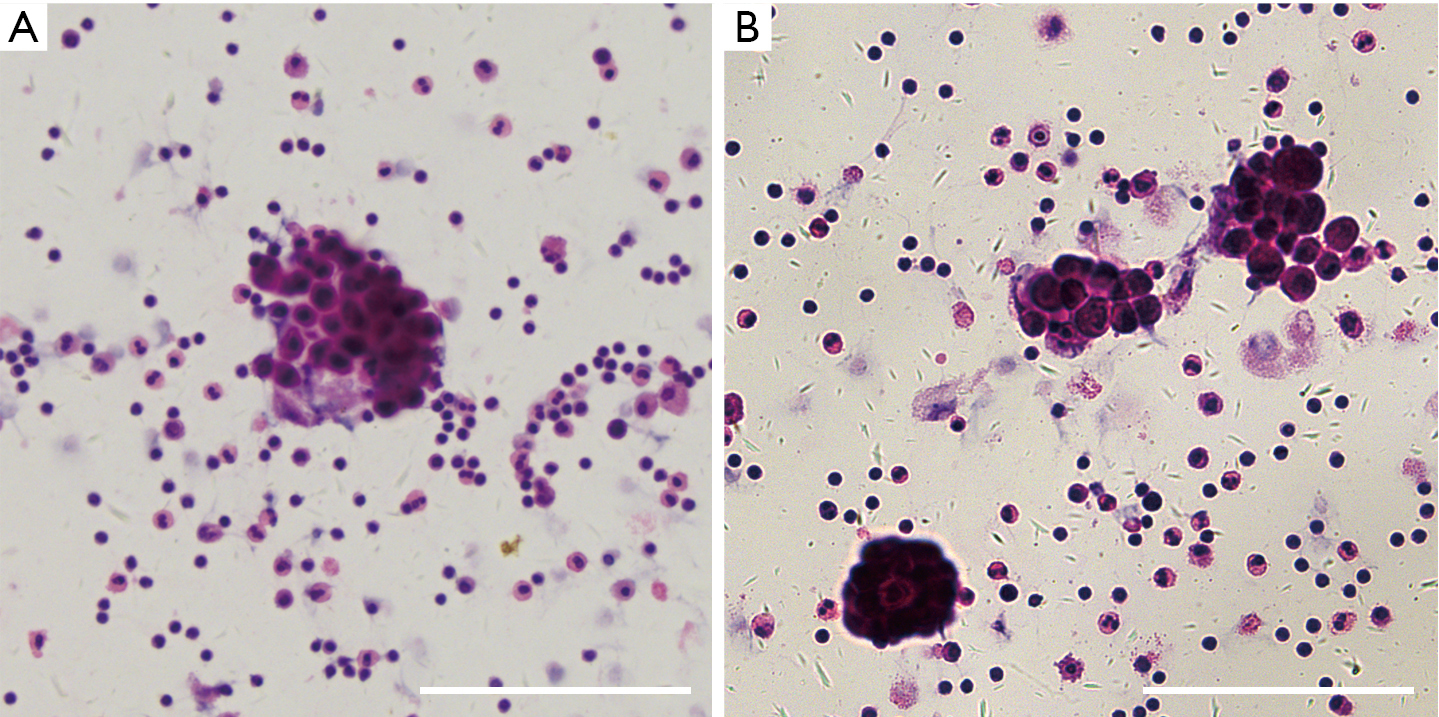
- A Cytocentrifugation Of Pleural Fluid Shows A Mixed Cell Population Download Scientific Diagram
- A Panel Of Markers For Identification Of Malignant And Non Malignant Cells In Culture From Effusions
- Benign Mesothelial Cells In Pleural Fluid Medical Laboratory Hematology Mad Scientist
- Pleural Mesothelioma Firefighters
- Free Printable I Love You Coloring Pages
- June Coloring Pages
- Cute Kawaii Coloring Pages For Kids
- Simple Flower Coloring Pages For Adults
If you re looking for Simple Flower Coloring Pages For Adults you've reached the ideal location. We ve got 104 images about simple flower coloring pages for adults including images, pictures, photos, backgrounds, and more. In such page, we also have number of graphics available. Such as png, jpg, animated gifs, pic art, symbol, blackandwhite, transparent, etc.
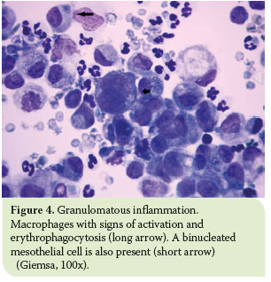
Hjcam Iatrikh Zwwn Syntrofias Hellenic Journal Of Companion Animal Medicine Volume 6 Issue 1 2017 Pleural Effusion In The Cat A Focus On Laboratory Diagnosis Simple Flower Coloring Pages For Adults
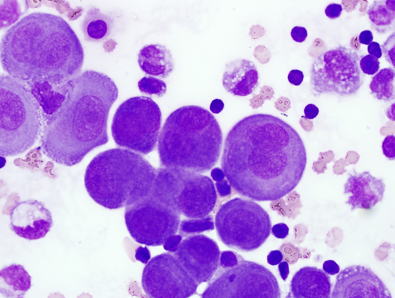
Reactive pleural effusion showing acute and chronic cells normal mesothelial cells and alveolar macrophages in.
Simple flower coloring pages for adults. Pleural mesothelial cells pmcs derived from the mesoderm play a key role during the development of the lung. A big step forward was the recent availability of the bap1 immunostain a relatively new marker that is expressed in the nuclei of benign mesothelial cells but is absent in greater than onehalf of cases of malignant mesothelioma. 17 the bap1 gene is located at 3p21 which is lost in approximately 30 of mesotheliomas.
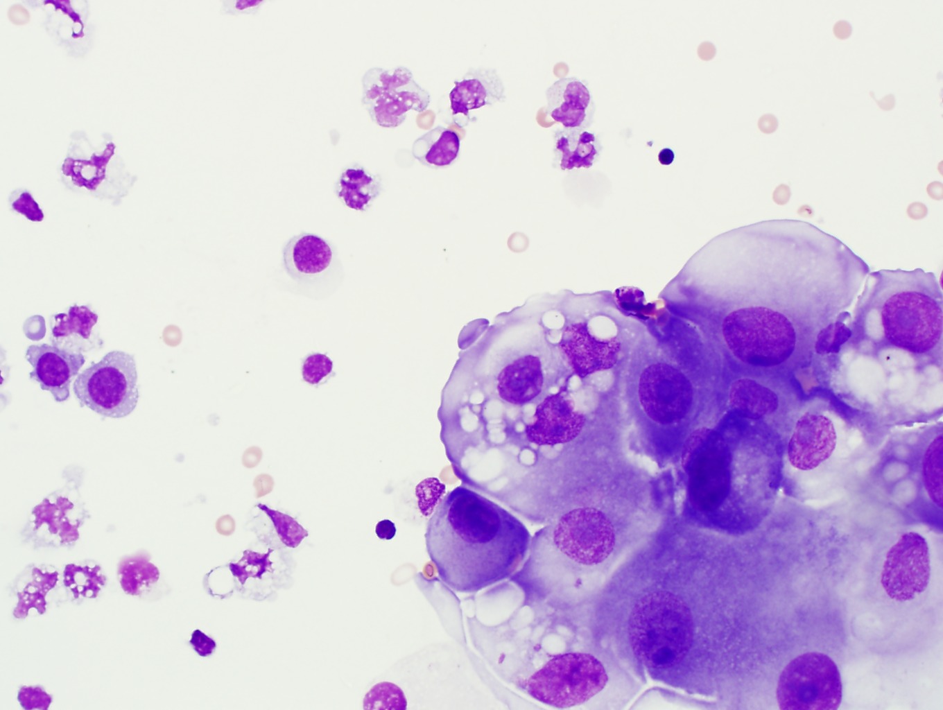
During development the mesoderm maintains a complex relationship with the developing endoderm giving rise to the mature lung. Pleural effusion showing monolayered sheet of normal mesothelial cells demonstrating spongiotic separation of individual cells within the sheet. A thoracentesis was performed and the pleural fluid was found to be straw colored and hazy with a red blood cell count of 1400mcl and a white blood cell count of 19 000 10 9 l.
Epithelial or lining cells most commonly mesothelial cells1 the appearance and presentation of nucleated cells found in pleural fluid and whether they are considered commonbenign or abnormal is discussed below. This course is intended for laboratory professionals who have experience with peripheral blood morphology and basic experience with body fluid differential analysisthis tutorial will provide a review of normal and abnormal body fluid morphology utilizing wright giemsa stained cytospin preparations from cerebrospinal fluid csf pleural peritoneal and synovial fluids as. Common cells present in pleural fluid include neutrophils lymphocytes monocytes mesothelial cells and red blood.
Cm 97 2003 pleural wright giemsa x320 identification referee participant lymphocyte reactive 607 411 plasma cell 214 183 mesothelial cell 107 112 malignant cell non heme 71 92 this photomicrograph is from the same case as cm 09 2003 above. The pleural mesothelium differentiates to give rise to the endothelium and smooth muscle cells via epithelial to mesenchymal transition emt. Papanicolaou x100 reactive pleural effusion.
Microscopic examination of the pleural fluid revealed mostly neutrophils and occasional reactive and large multinucleated mesothelial cells demonstrating active. Also seen in this field are a plasma cell a macrophage and a neutrophil. Mesothelial cells in pleural fluid eighty five samples of mesothelial cells in pleural fluid from 76 patients with biopsy proven tuberculous pleurisy were examined cytologically.
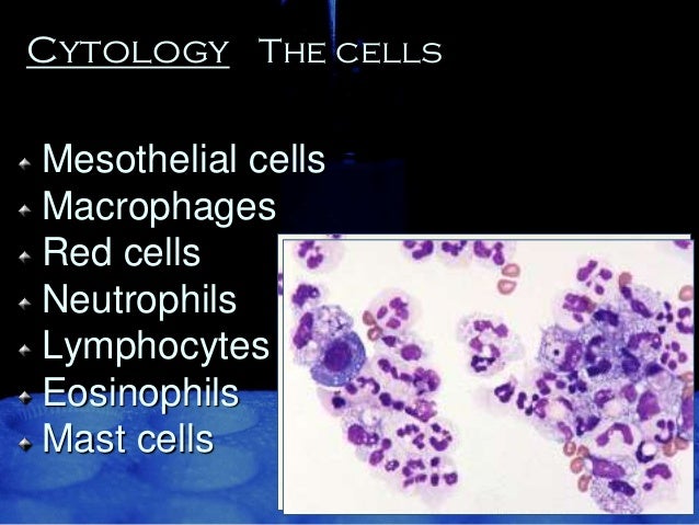
A Cytocentrifugation Of Pleural Fluid Shows A Mixed Cell Population Download Scientific Diagram Simple Flower Coloring Pages For Adults
More From Simple Flower Coloring Pages For Adults
- Flamingo Coloring Pages Pdf
- Race Car Coloring Pages Free
- Personal Attorney Near Me
- Mega Evolution Charizard Coloring Page
- Kahlon Law Office
Incoming Search Terms:
- Effusions Abdominal Thoracic And Pericardial Veterian Key Kahlon Law Office,
- Http Www Api Pt Com Reference Commentary 2015ascope Pdf Kahlon Law Office,
- Effusions Cytopathology Cellnetpathology Kahlon Law Office,
- Https Link Springer Com Content Pdf 10 1007 2f978 94 009 0849 9 3 Pdf Kahlon Law Office,
- Http Www Api Pt Com Reference Commentary 2015ascope Pdf Kahlon Law Office,
- Fluid Cytology In Serous Cavity Effusions Kahlon Law Office,

