Mesothelial Cells In Pleural Fluid, Pleural Fluid Mast Cells 5
Mesothelial cells in pleural fluid Indeed lately is being sought by consumers around us, maybe one of you personally. Individuals now are accustomed to using the internet in gadgets to see image and video data for inspiration, and according to the title of the article I will discuss about Mesothelial Cells In Pleural Fluid.
- Mesothelial Cells Continued Labce Com Laboratory Continuing Education Cell Body Fluid Continuing Education
- Cytology Of Pleural And Peritoneal Lesions Chapter 5 Practical Pathology Of Serous Membranes
- Mesothelial Cell Stock Photo Download Image Now Istock
- Home
- Effusions Abdominal Thoracic And Pericardial Veterian Key
- Figure 3 From Telomere Repeat Amplification Protocol Trap In Situ Reveals Telomerase Activity In Three Cell Types In Effusions Malignant Cells Proliferative Mesothelial Cells And Lymphocytes Semantic Scholar
Find, Read, And Discover Mesothelial Cells In Pleural Fluid, Such Us:
- Differentiation Of Mesothelial Cells Into Macrophage Phagocytic Cells In A Patient With Clinical Sepsis
- Home
- Pleural Fluid For Cytology Dr Sachin Kale
- J C Prolla Cytopathology Ascites Pancreatitis Mesothelial Cell Atypias
- Https Encrypted Tbn0 Gstatic Com Images Q Tbn 3aand9gcseb7aizju0qbedevvi8irefmeqt7 Rh3pa 8irkz1p4jn0mu35 Usqp Cau
- Carrabelle Restaurant Dayton Tn
- Dog Coloring Pages Free
- John Langford Law
- Vision Law
- Cute Cloud Coloring Page
If you are looking for Cute Cloud Coloring Page you've come to the ideal place. We ve got 104 images about cute cloud coloring page including images, photos, pictures, wallpapers, and more. In such web page, we additionally provide variety of graphics available. Such as png, jpg, animated gifs, pic art, symbol, black and white, translucent, etc.
We demonstrated that pleural fluid post lung surgery is a source of mesothelial.

Cute cloud coloring page. The broad expression of cd71 molecule in postoperative pleural fluid suggests that many of the free floating non leukocyte cells were activated or proliferative mesothelial cells. The pleural mesothelial cell pmc is the most common cell in the pleural space and is the primary cell that initiates responses to noxious stimuli. Pmcs are metabolically active cells that maintain a dynamic state of homeostasis in the pleural space.
Of 31 exudative effusions with a lymphocytic predominance 30 were due either to tuberculosis or neoplasm. No significant differences have been identified between the mesothelial cells in the visceral or parietal pleura or among those in the pleural peritoneal or pericardial cavities. In contrast 653 of pleural fluid aspirates obtained from a control group of pati.
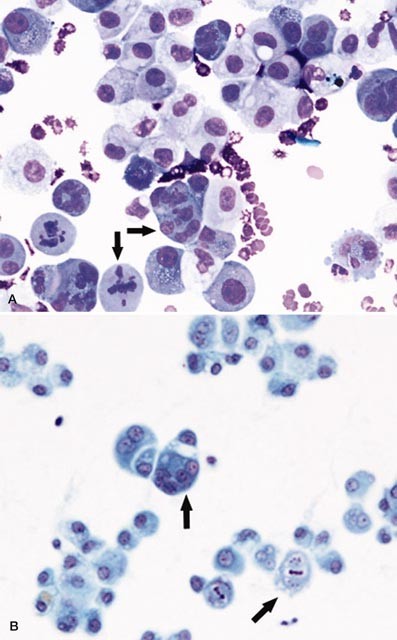
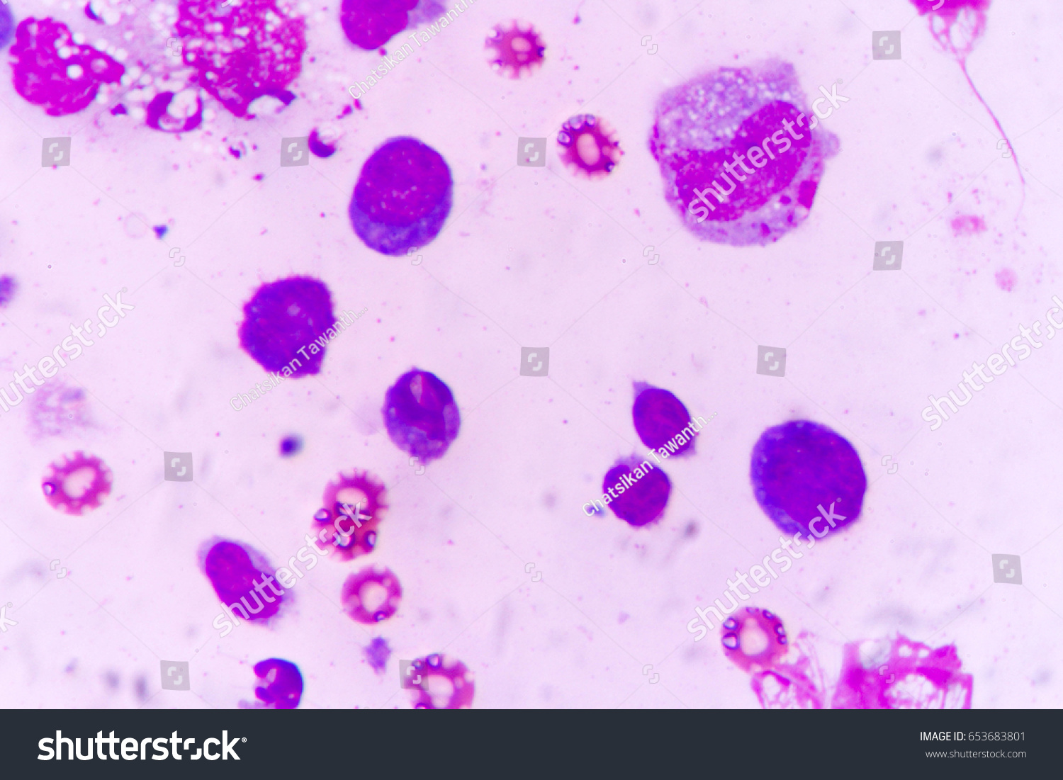
Mesothelial cell is the predominant cell type in the pleural cavity see mesothelial cells. The mesothelial cells have central round nuclei with a moderate amount of light purple cytoplasm and a corona or fringe to the cytoplasmic borders. Mesothelial cells in pleural fluid.
Eighty five samples of pleural fluid obtained from 76 patients with biopsy proven tuberculous pleurisy were examined cytologically. Use of pleural fluid n. Pleural fluid cytological studies showed malignant cells in 33 of 43 patients with effusions due to tumor.
Numerous mesothelial cells are seen in this pleural fluid from a dog with a transudative effusion with concurrent diapedesis of red blood cells or hemorrhage. Pleural effusions are a build up of fluid in the cavity between the two layers of the pleura the pleural mesothelium and is influenced by substances secreted by pleural mesothelial cells. Mesothelial cells are found in variable numbers in most effusions but their presence at greater than 5 of total nucleated cells makes a diagnosis of tb less likely.
Numerous reactive mesothelial cells were present in only 12 of specimens examined. There are many possible causes of pleural effusions.
More From Cute Cloud Coloring Page
- Can Mesothelioma Be Diagnosed
- Mesothelioma Tonofilaments
- Tax Attorney Meme
- Lawrence Lawrence
- Scary Dark Images
Incoming Search Terms:
- Cytology Of Pleural Fluid Clumps Of Neoplastic Cells With Download Scientific Diagram Scary Dark Images,
- Malignant Cells In Pleural Fluid Stock Photo Image Of Science Healthcare 98411708 Scary Dark Images,
- Atypical Mesothelial Cells In Pleural Effusion Youtube Scary Dark Images,
- Effusions Abdominal Thoracic And Pericardial Veterian Key Scary Dark Images,
- Cytology Of Pleural And Peritoneal Lesions Chapter 5 Practical Pathology Of Serous Membranes Scary Dark Images,
- Differentiation Of Mesothelial Cells Into Macrophage Phagocytic Cells In A Patient With Clinical Sepsis Scary Dark Images,


