Reactive Mesothelial Cells, Immunohistochemical Differentiation Of Reactive From Malignant Mesothelium As A Diagnostic Aid In Canine Pericardial Disease Milne 2018 Journal Of Small Animal Practice Wiley Online Library
Reactive mesothelial cells Indeed recently is being sought by users around us, maybe one of you personally. People are now accustomed to using the internet in gadgets to see video and image data for inspiration, and according to the title of this article I will discuss about Reactive Mesothelial Cells.
- Fluid Cytology In Serous Cavity Effusions
- Http Jtd Amegroups Com Article Viewfile 25454 Pdf
- Pathology Outlines Mesothelial
- The Don T Eat Me Signal Cd47 Is A Novel Diagnostic Biomarker And Potential Therapeutic Target For Diffuse Malignant Mesothelioma Abstract Europe Pmc
- Sheet Of Reactive Mesothelial Cells Note Windows Between Cells And Download Scientific Diagram
- Pb Reactive Mesothelial Hyperplasia Mimicking Mesothelioma In An African Green Monkey Chlorocebus Aethiops
Find, Read, And Discover Reactive Mesothelial Cells, Such Us:
- Mesothelial Cytopathology Libre Pathology
- Fluid Cytology In Serous Cavity Effusions
- Pleural Fluid Clusters Of Atypical Reactive Mesothelial Cells From A Download Scientific Diagram
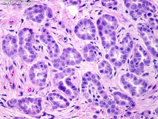
- The Cell Morphological Characteristics Of Adenocarcinoma Cells Download Scientific Diagram
- Peritoneal Fluid Having Reactive Mesothelial Cells As Well As Mixed Download Scientific Diagram
- Memphis Personal Injury Lawyer
- Grinch Pictures To Print
- Stephen Meyer Quotes
- Mesothelioma Law Firm U
- Giraffe Coloring Pages For Adults
If you re searching for Giraffe Coloring Pages For Adults you've reached the right place. We ve got 104 images about giraffe coloring pages for adults including images, pictures, photos, wallpapers, and more. In these web page, we also have variety of graphics out there. Such as png, jpg, animated gifs, pic art, symbol, black and white, translucent, etc.
Home Giraffe Coloring Pages For Adults
Pb Reactive Mesothelial Hyperplasia Mimicking Mesothelioma In An African Green Monkey Chlorocebus Aethiops Giraffe Coloring Pages For Adults
J C Prolla Cytopathology Ascites Pancreatitis Mesothelial Cell Atypias Giraffe Coloring Pages For Adults
Webpathology Com A Collection Of Surgical Pathology Images Giraffe Coloring Pages For Adults

The Panorama Of Different Faces Of Mesothelial Cells Basicmedical Key Giraffe Coloring Pages For Adults

Pdf A Cytological Study To Differentiate Between Reactive Mesothelial Cells And Malignant Cells In Effusions Semantic Scholar Giraffe Coloring Pages For Adults
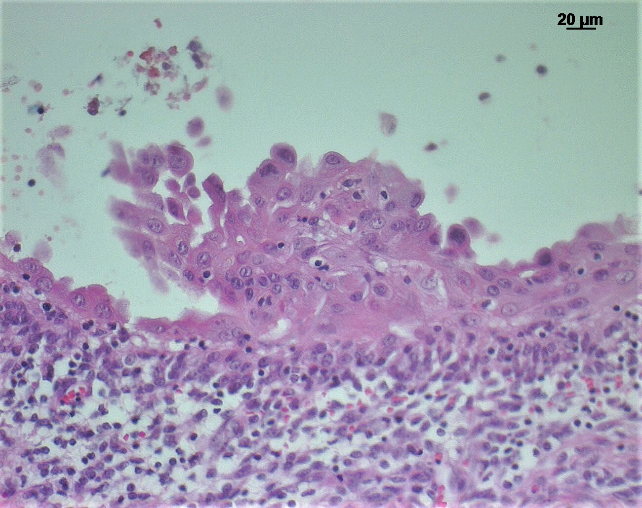
The healing of disrupted serosa includes the multiplication and migration of mesothelial cells from the edges of the injured area.
Giraffe coloring pages for adults. Weiss in modern surgical pathology second edition 2009. The main purpose of these cells is to produce a lubricating fluid that is released between layers providing a slippery non adhesive and protective surface to facilitate intracoelomic movement. Materials and methods.
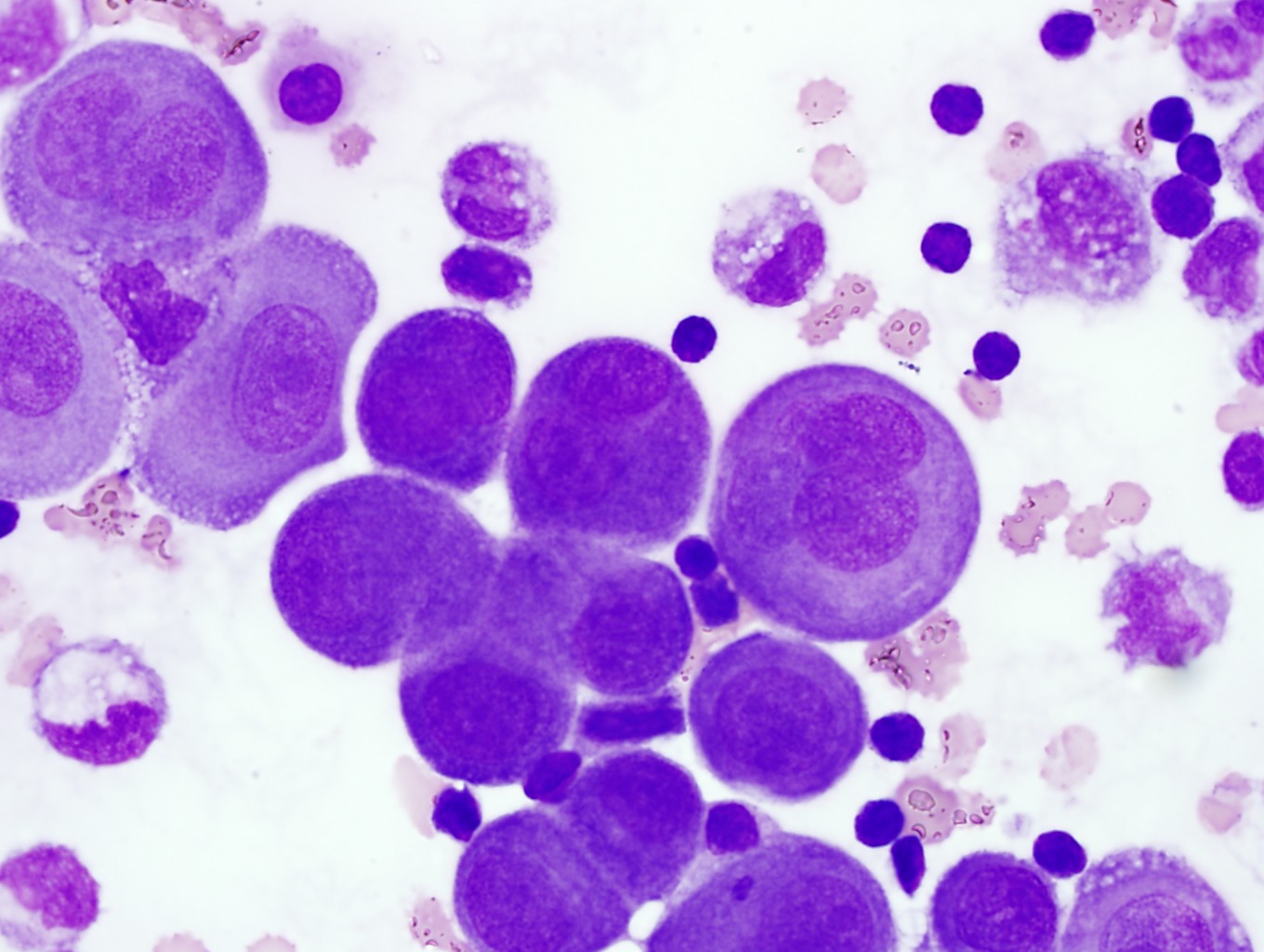
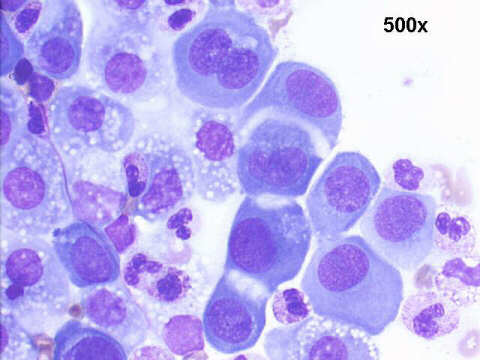
It can also be the result of trauma or the presence of metastatic tumor. The mesothelium is composed of an extensive monolayer of specialized cells mesothelial cells that line the bodys serous cavities and internal organs. Benign mesothelial cells tend to arrange in monolayered sheets with little nuclear overlapping fig.
D whitaker d shilkin k. Desmin and neural marker expression in mesothelial cells and mesotheliomas. Please note the image on the right the cytoplasmic borders are clear in this group compared with normal mesothelial cells.
Reactive mesothelial cells may sometimes be sampled in mediastinal fine needle aspirates and should not be mistaken as thymoma cells. Under normal conditions mesothelial cells form a flat single uniform layer. The use of.
This condition can be due to the presence of a bacterial viral or fungal infection. Reactive mesothelial cells tend to come in clusters and patches and are more faded in the cytoplasm of body fluids. Various immunocytochemical icc markers have been used to maximize the diagnostic accuracy however cytopathologists still e.
It was an observational studyconducted in the. There are certain cells that line the pleura the thin double layered lining which covers the lungs chest wall and diaphragm which are known as mesothelial cellsother than the pleura mesothelial cells also form a lining around the heart pericardium and the internal surface of the abdomen peritoneum. Reactive mesothelial cells hence will help in making an early and accurate diagnosis.
J pathol 1993169sl188a abstract. Mesothelial cells in pleural fluid. It may also include repopulation from free floating mesothelial cells or possibly.
Mesothelial cells are mesodermally derived epithelial cells that line body cavities pleura pericardium and peritoneum. Separation of nuclei large and well defined nucleoli help identify corrective mesothelial cells. When these surfaces become irritated or injured mesothelial cells can proliferate and take on a variety of morphologic and cytologic appearances.
They contain ovoid nuclei fine chromatin delicate nuclear membrane small nucleoli and a moderate. Expression of desmin and smooth muscle actin in mesothelial hyperplasia and mesothelioma. Mesothelial hyperplasia represents a normal reaction to injury.
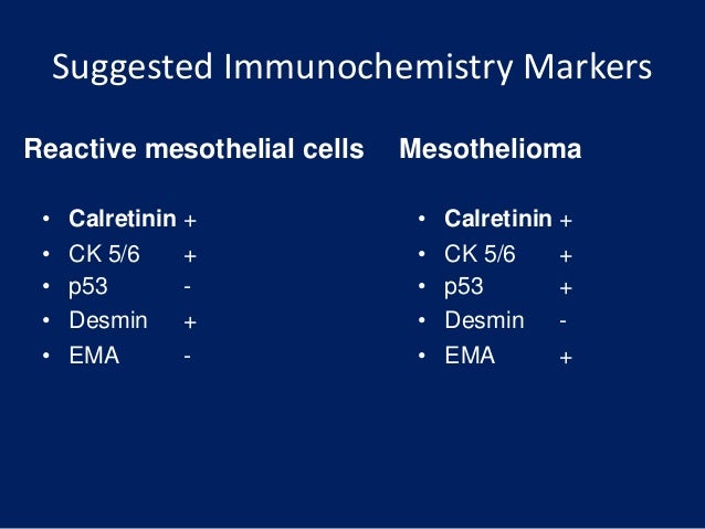
To differentiate reactive mesothelial cells and adenocarcinoma cells using immunocytochemical markers ber ep 4 moc 31 calretinin and hbme 1 in serous effusions.
Fluid Cytology In Serous Cavity Effusions Giraffe Coloring Pages For Adults
Mesothelial Cytopathology Libre Pathology Giraffe Coloring Pages For Adults

Non Neoplastic Reactive Mesothelial Cells Dog With Non Neoplastic Download Scientific Diagram Giraffe Coloring Pages For Adults
Https Biomedres Us Pdfs Bjstr Ms Id 003107 Pdf Giraffe Coloring Pages For Adults
More From Giraffe Coloring Pages For Adults
- Chinese Law Firm
- Steve Harris Law And Order
- Dolphin Coloring Pages
- Idiopathic Peritoneal Mesothelioma
- Mesothelioma Staging Radiology
Incoming Search Terms:
- Reactive Mesothelial Cells Can Exhibit Coarse Chromatin Irregular Nuclear Outlines And Nucleoli It Is Important To Microscopic Cells Medical Laboratory Cell Mesothelioma Staging Radiology,
- Figure 1 From Cytological Evaluation Of Serous Body Fluids A Two Year Experience In Tertiary Care Centre From Central India Semantic Scholar Mesothelioma Staging Radiology,
- Signet Ring Cell Reactive Mesothelial Cell Stock Photo Edit Now 1260129097 Mesothelioma Staging Radiology,
- Reactive Mesothelial Cells Hematology Medical Laboratory Science Medical Laboratory Mesothelioma Staging Radiology,
- Reactive Mesothelial Cell Paracentesis Ascities 100x Giemsa Stain Stain Microscopic Mesothelioma Staging Radiology,
- Benign Elements Eurocytology Mesothelioma Staging Radiology,
