Reactive Mesothelial Cells In Ascitic Fluid, International Journal Of Current Research And Review Ijcrr
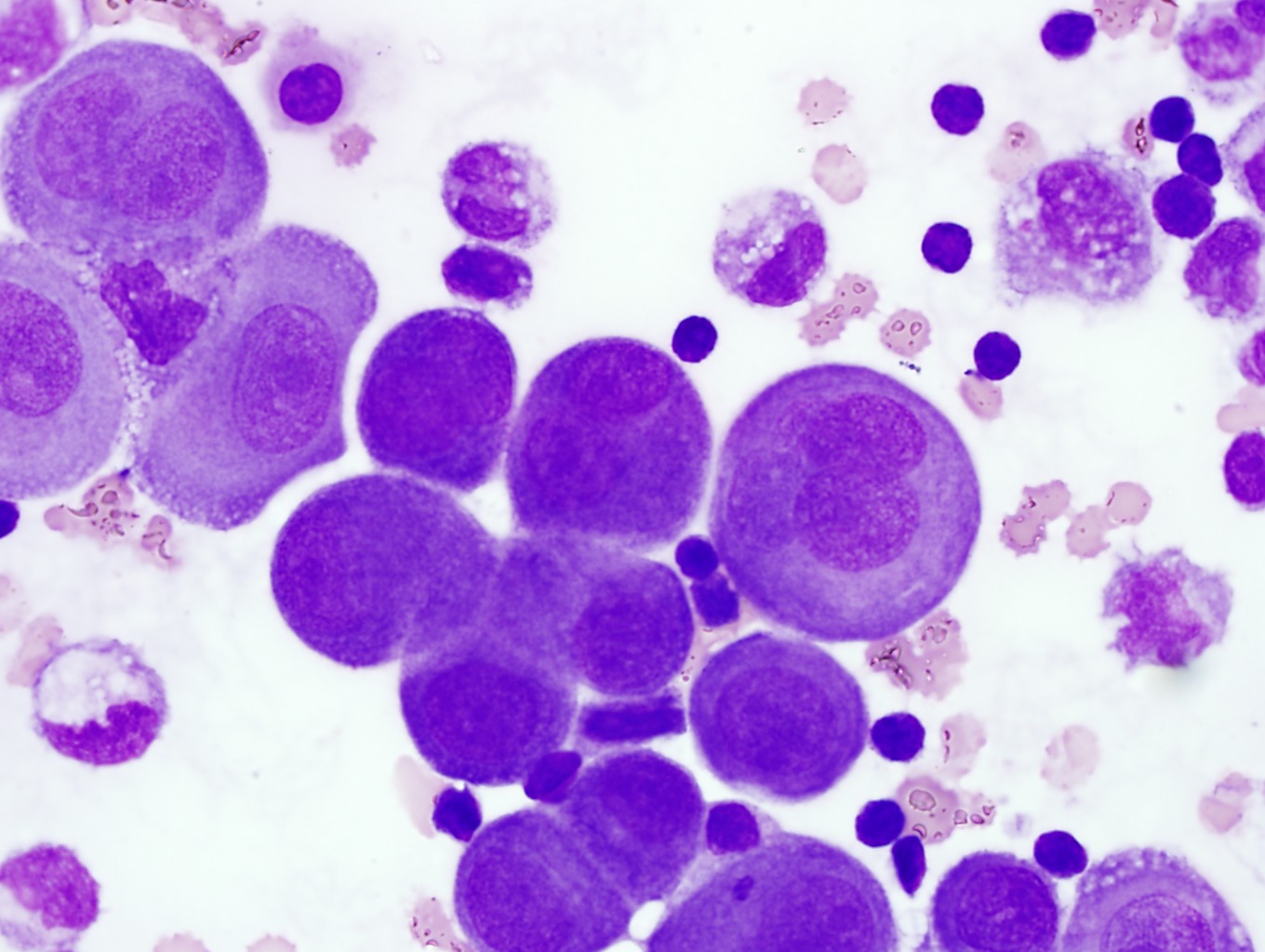
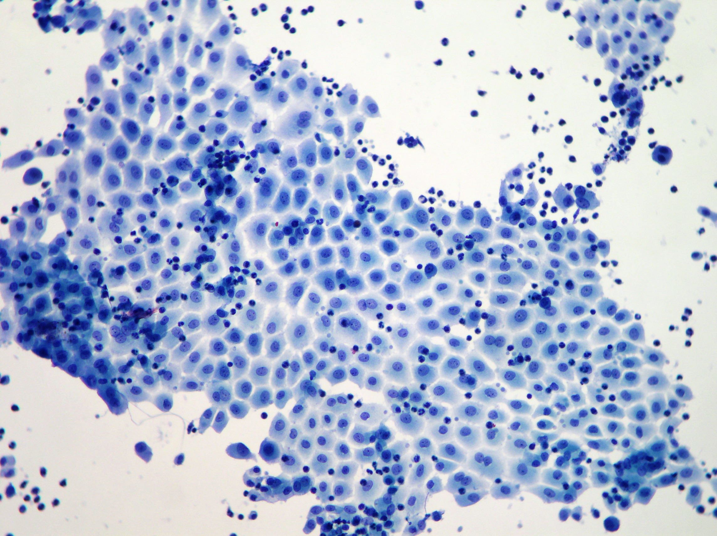
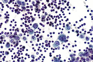
Reactive mesothelial cells in ascitic fluid Indeed recently is being hunted by consumers around us, maybe one of you personally. People now are accustomed to using the net in gadgets to view video and image information for inspiration, and according to the name of the article I will discuss about Reactive Mesothelial Cells In Ascitic Fluid.
- Cytology Of Ascitic Fluid A The Smear Is Moderately Cellular With Download Scientific Diagram
- Https Www Seap Es Documents 228448 526984 03 Jorda Pdf
- Home
- Http Iap Ad Org Documents Archieve Congress 25thcongress Workshop First 20day Body 20fluids 20cytology Pdf
- Significance Of Flower Pot Cells In Effusion Cytology Bharani 2017 Diagnostic Cytopathology Wiley Online Library
- J C Prolla Cytopathology Ascites Pancreatitis Mesothelial Cell Atypias
Find, Read, And Discover Reactive Mesothelial Cells In Ascitic Fluid, Such Us:
- Home
- Https Www Rcpath Org Asset Ed8cdd8d 8d04 4b82 Ad48d585e2f023be
- Mesothelial Hyperplasia An Overview Sciencedirect Topics
- Ppt Analysis Of Body Cavity Fluids Powerpoint Presentation Free Download Id 4125931
- Https Www Seap Es Documents 228448 526984 03 Jorda Pdf
- Disney Coloring Pages For Adults Easy
- Chrysotile Insulation
- Spectrum Nyc Fs1
- Chippewa Township Mesothelioma Lawyers
- Child And Family Law
If you are looking for Child And Family Law you've arrived at the right location. We have 104 graphics about child and family law adding pictures, pictures, photos, backgrounds, and more. In such webpage, we additionally provide number of images out there. Such as png, jpg, animated gifs, pic art, logo, black and white, transparent, etc.
A 55 year old male presented with gradually progressive ascites.

Child and family law. Ascitic fluid analysis or peritoneal fluid analysis is the major diagnostic test to study the pathophysiology of accumulation of fluid in the peritoneum including diagnosing the causes and inflammation of the fluids. Reactive mesothelial cells can be found when there is an infection or an inflammatory response present in a body cavity. There is chromatin clumping and changed the nuclearcytoplasmic ratio.
The ascitic fluid is aspirated from the peritoneal cavity. This condition can be due to the presence of a bacterial viral or fungal infection. It can also be the result of trauma or the presence of metastatic tumor.
In inflammatory conditions there is a greater number of reactive mesothelial and polys whereas in case of transudate there. The reactive mesothelial proliferations form smaller uniform less complex groups compared to malignant mesothelial proliferations. Carlos w m bedrossian discusses at length the great variability of mesothelial cells.
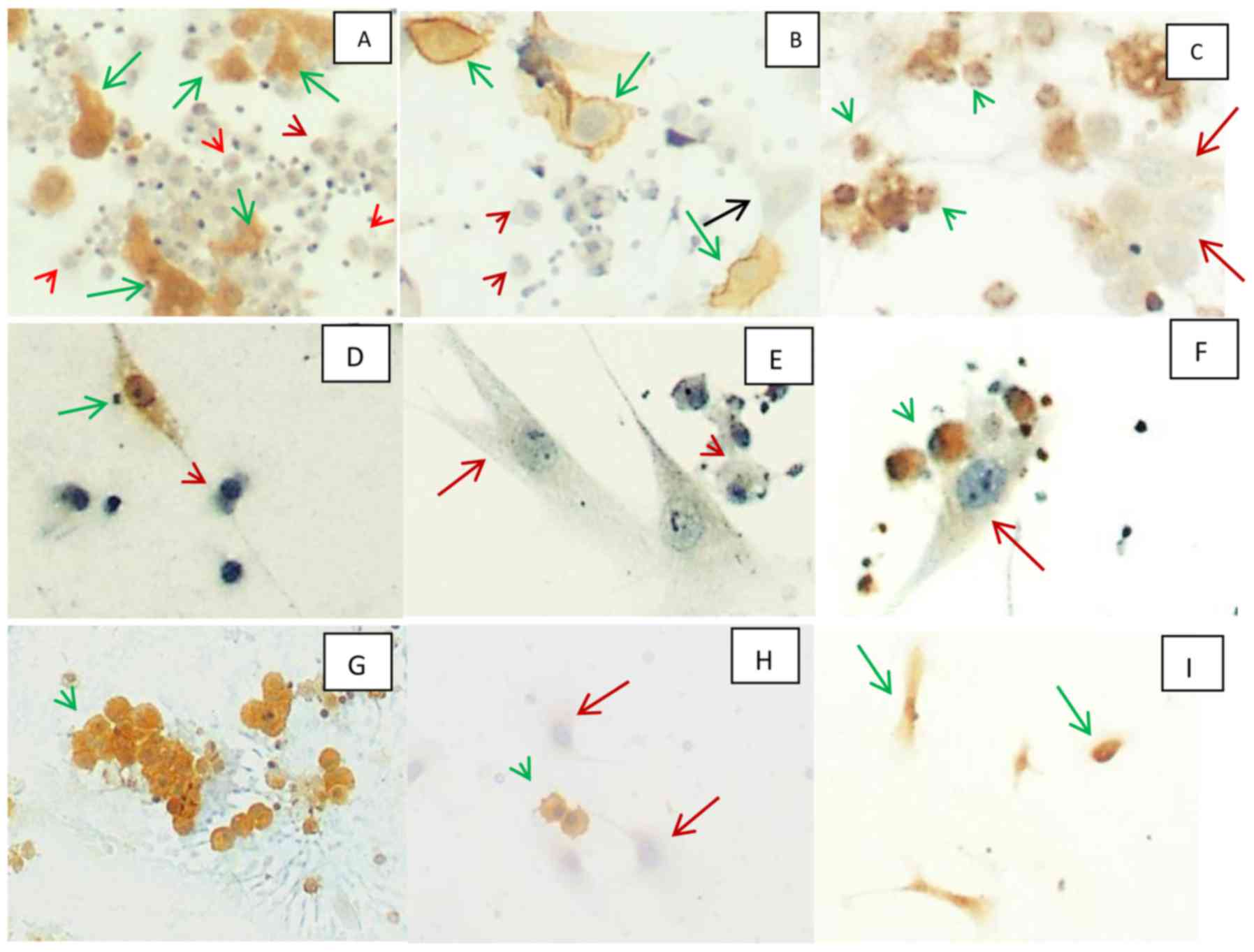
Further work up the tumor cells were also positive for wt1 calretinin figure 1c1 and andc2 c2 p53 and cytokeratin 56 and stained negatively for pax 8 berep4 desmin cea and ttf 1. Some time reactive mesothelial cells and malignant cell differentiation is difficult. Cytospin preparations from ascitic fluid showed reactive mesothelial cells admixed with few smooth contoured clusters of cells with moderate cytoplasm vesicular nuclei with prominent nucleolus.
Serosal reaction to injury p26 51 dr. Malignant cells have the variable morphology of the cells and nuclei.
More From Child And Family Law
- Mesothelioma Catheter
- Rainbow Coloring Worksheet
- Mesothelioma In The Mouth
- Mental Health Attorney New York
- Real Estate Law Firms Miami
Incoming Search Terms:
- Full Text Cytological Diagnosis Of Metastatic Deposits Of Alveolar Rhabdomyosarcoma In Ascitic Fluid A Rare Case Report Edorium Journal Of Oncocytology Real Estate Law Firms Miami,
- Http Www Asl5 Liguria It Portals 0 Anatomiapatologica2015 20150924 Effusion Cytology Pdf Real Estate Law Firms Miami,
- Https Encrypted Tbn0 Gstatic Com Images Q Tbn 3aand9gctjxi34atmipfik6awy9fwku9hnngvwavizoylai85qzyd 85bh Usqp Cau Real Estate Law Firms Miami,
- Serous Effusions Basicmedical Key Real Estate Law Firms Miami,
- Effusion Cytology Clinician S Brief Real Estate Law Firms Miami,
- Diagnostic Pitfalls In Effusion Fluid Cytology Basicmedical Key Real Estate Law Firms Miami,


