Reactive Mesothelial Cells Vs Mesothelioma, Https Academic Oup Com Ajcp Article Pdf 110 3 397 24884795 Ajcpath110 0397 Pdf
Reactive mesothelial cells vs mesothelioma Indeed lately has been sought by users around us, maybe one of you personally. Individuals now are accustomed to using the internet in gadgets to view image and video data for inspiration, and according to the title of this article I will discuss about Reactive Mesothelial Cells Vs Mesothelioma.
- Common Immunohistochemical Stains Used To Differentiate Pulmonary Download Table
- Benign And Malignant Mesothelial Proliferation Semantic Scholar
- 02 Presentations Ii Vs 14 4 Mb 3 30 08
- Utility Of Cell Block To Detect Malignancy In Fluid Cytology Adjunct Or Necessity Dey S Nag D Nandi A Bandyopadhyay R J Can Res Ther
- Swathi Prabhu Md On Twitter How To Differentiate Reactive Mesothelial Cells From Mesothelioma Dr Radhika Srinivasan Cytopathology Cytopath Kapcon2020 Pathridle Susan Vi Monappa Drramaswamyas Anjuthevirgo Drsupriyatiwari Drgeeone Https T
- Http Handouts Uscap Org 2016 Cm06 Daci 1 Pdf
Find, Read, And Discover Reactive Mesothelial Cells Vs Mesothelioma, Such Us:
- Malignant Mesothelioma A Histomorphological And Immunohistochemical Study Of 24 Cases From A Tertiary Care Hospital In Southern India Hui M Uppin Sg Bhaskar K Kumar Nn Paramjyothi Gk Indian J Cancer
- Https Academic Oup Com Ajcp Article Pdf 110 3 397 24884795 Ajcpath110 0397 Pdf
- Gale Academic Onefile Document Utility Of Survivin Bap1 And Ki 67 Immunohistochemistry In Distinguishing Epithelioid Mesothelioma From Reactive Mesothelial Hyperplasia
- Https Academic Oup Com Ajcp Article Pdf 110 3 397 24884795 Ajcpath110 0397 Pdf
- Problems In Mesothelioma Diagnosis Addis 2009 Histopathology Wiley Online Library
- Scary Pumpkin
- Realistic Bee Coloring Pages
- Rocket Ship Coloring Pages Printable
- Grey Asbestos Floor Tiles
- Mesothelioma Asbestos Class Action
If you are looking for Mesothelioma Asbestos Class Action you've arrived at the perfect place. We have 104 graphics about mesothelioma asbestos class action including pictures, photos, pictures, backgrounds, and much more. In these page, we also have variety of images out there. Such as png, jpg, animated gifs, pic art, symbol, blackandwhite, translucent, etc.
This condition can be due to bacteria virus or fungus.
Mesothelioma asbestos class action. Archival paraffin embedded cell blocks of pleural and peritoneal fluids from 52 patients with malignant mesothelioma mm and 64 patients with reactive mesothelial hyperplasia mh were retrieved. 163299 305 26 kato y tsuta k seki k et al. The distinction between reactive mesothelial hyperplasia mh and malignant mesothelioma mm may be very difficult based only on histologic and morphologic findings.
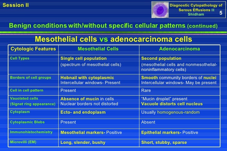
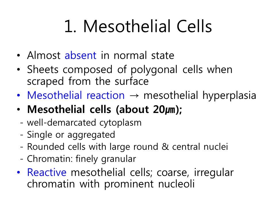
Reactive mesothelial cells mesothelioma architecture flat sheets 3 d businesses organization size. The cells show intense cytoplasmic staining. B immunostaining of rms for ecadherin.
This study tested the hypothesis that immunocytochemistry ic in effusion cell blocks cb can distinguish mm from rm and that the results may be applied to individual specimens. Mesothelial cytopathology is a huge part of cytopathology. Immunohistochemical detection of glut 1 can discriminate between reactive mesothelium and malignant mesothelioma.
Mesothelioma is a cancer of the lung linings that contain such cells. C immunostaining of rms for calretinin. Layeld cd147 immunohistochemistry discriminates between reactive mesothelial cells and malignant.
It also can be the result of trauma or a tumor. Reactive mesothelial cells vs mesothelioma. P16 fish followed by immunofluorescence with ema was helpful towards identifying the mesothelioma cells in the cell blocks.
Confidence in the diagnosis is often proportional to the amount of tissue available for study and depends largely on findings of invasion and t. In biopsy tissue discrimination between reactive mesothelial hyperplasia and epithelial mesothelioma can pose a major problem for the surgical pathologist. A papanicolaou staining of reactive mesothelial cells rms.
Note complete absence of staining. Symposium issue guest editor lester j. Distinguishing malignant mesothelioma mm from reactive mesothelial hyperplasia rm may be difficult in effusions.
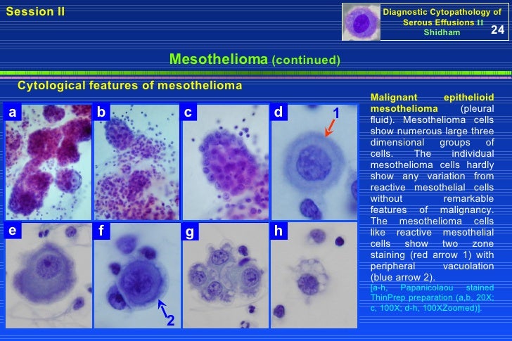
Reactive mesothelial cells are found when there is an infection or some type of inflammatory response in the body. Seventeen of the 22 mesothelioma patients 773 showed homozygous deletions of p16 in the tumor tissue and in the atypical mesothelial cells from the cell blocks. D papanicolaou staining of malignant mesothelioma mm cells.
E immunostaining of mm cells for ecadherin. Ihc stains included desmin epithelial membrane antigen ema glucose transport protein 1 glut 1 ki67 and p53.
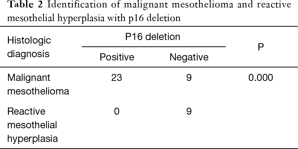
Role Of P16 Deletion And Bap1 Loss In The Diagnosis Of Malignant Mesothelioma Liu Journal Of Thoracic Disease Mesothelioma Asbestos Class Action
More From Mesothelioma Asbestos Class Action
- Neoadjuvant Therapy Mesothelioma
- Harris Law
- Little Girl Coloring Pages Printable
- Pokemon Coloring Pages Eevee Evolutions
- What Famous Actor Set Up A Mesothelioma Trust Fund
Incoming Search Terms:
- Webpathology Com A Collection Of Surgical Pathology Images What Famous Actor Set Up A Mesothelioma Trust Fund,
- Reactive Mesothelial Hyperplasia Springerlink What Famous Actor Set Up A Mesothelioma Trust Fund,
- Webpathology Com A Collection Of Surgical Pathology Images What Famous Actor Set Up A Mesothelioma Trust Fund,
- Mesothelial Cytopathology Libre Pathology What Famous Actor Set Up A Mesothelioma Trust Fund,
- Https Www Rcpath Org Asset Ed8cdd8d 8d04 4b82 Ad48d585e2f023be What Famous Actor Set Up A Mesothelioma Trust Fund,
- Https Www Hkiap Org Wp Content Uploads Lecture Notes 2019 20special 20cytology 20workshop Mesothelioma 20how 20far Pdf What Famous Actor Set Up A Mesothelioma Trust Fund,
