Mesothelioma Vs Reactive Mesothelial Cells Cytology, Reactive Mesothelial Hyperplasia Springerlink
Mesothelioma vs reactive mesothelial cells cytology Indeed recently is being sought by consumers around us, maybe one of you. Individuals are now accustomed to using the net in gadgets to see image and video information for inspiration, and according to the title of the article I will talk about about Mesothelioma Vs Reactive Mesothelial Cells Cytology.
- Https Encrypted Tbn0 Gstatic Com Images Q Tbn 3aand9gcqhd7waupdnvkltywmzp1zjlgtbh2fsowenirnocak8jd8 Jgve Usqp Cau
- Cytologic Differential Diagnosis Among Reactive Mesothelial Cells Malignant Mesothelioma And Adenocarcinoma Kitazume 2000 Cancer Cytopathology Wiley Online Library
- Effusion Cytology Clinician S Brief
- Malignant And Borderline Mesothelial Tumors Of The Pleura Sciencedirect
- Https Www Rcpath Org Asset Ed8cdd8d 8d04 4b82 Ad48d585e2f023be
- Reactive Mesothelial Hyperplasia Springerlink
Find, Read, And Discover Mesothelioma Vs Reactive Mesothelial Cells Cytology, Such Us:
- Reactive Mesothelial Hyperplasia Springerlink
- Reactive Mesothelial Hyperplasia Springerlink
- Cytological Diagnosis Of Mesothelioma Dr Sampurna Roy Md
- Https Encrypted Tbn0 Gstatic Com Images Q Tbn 3aand9gcrgrvn 3be6wylpxdte0su7bvf9lduonoausevalghb5uds Xu6 Usqp Cau
- Utility Of Survivin Bap1 And Ki 67 Immunohistochemistry In Distinguishing Epithelioid Mesothelioma From Reactive Mesothelial Hyperplasia
- Scary Woods
- Gwendolyn Knight Lawrence
- Squamous Cell Carcinoma Lung Xray
- Monster High Coloring Pages
- Sports Coloring Pages For Girls
If you are looking for Sports Coloring Pages For Girls you've reached the perfect location. We ve got 104 graphics about sports coloring pages for girls including pictures, photos, photographs, backgrounds, and much more. In such page, we additionally provide variety of graphics out there. Such as png, jpg, animated gifs, pic art, symbol, black and white, transparent, etc.
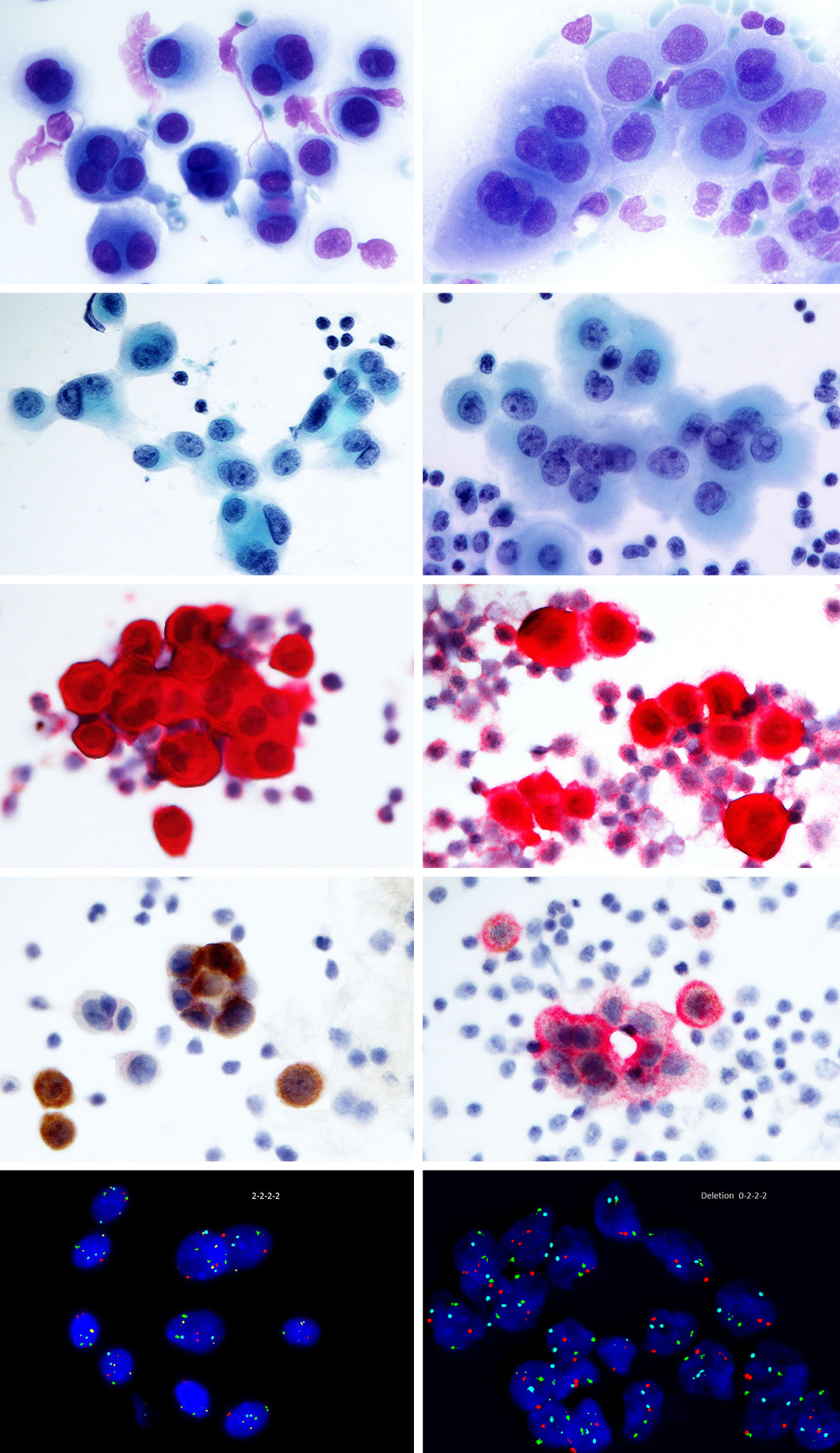
Malignant mesothelial cells show strong and diffuse positivity.

Sports coloring pages for girls. Most adenocarcinoma smears showed a pop. Immunohistochemical detection of glut 1 can discriminate between reactive mesothelium and malignant mesothelioma. Mesothelioma patients often present with serosal effusions which are ideal for cytopathological diagnoses.
The differential diagnosis between reactive mesothelial cells rms malignant mesotheliomas mms and adenocarcinomas acs is often difficult in cytologic specimens and the utility of various immunohistochemical markers have been explored. Confidence in the diagnosis is often proportional to the amount of tissue available for study and depends largely on findings of invasion and t. Xiap was not a sensitive marker for malignancy and had a limited value in cytology.
In biopsy tissue discrimination between reactive mesothelial hyperplasia and epithelial mesothelioma can pose a major problem for the surgical pathologist. Compared with calretinin d2 40 was a more sensitive marker of mesothelial cells. Moc 31 and d2 40 were very sensitive and specific markers of epithelial and mesothelial cells respectively.
163299 305 26 kato y tsuta k seki k et al. The differential diagnosis of epithelial type mesothelioma from adenocarcinoma and reactive mesothelial proliferation. Malignant mesothelial cells show strong membranous positivity with some cytoplasmic staining.
However the morphological overlap between malignant and benign mesothelial proliferation can make a conclusive cytological diagnosis of mesothelioma elusive because immunohistochemical staining does not discriminate definitively between the two in this setting. Reactive mesothelial cells show negative to weak positivity. Mesothelioma vs reactive mesothelial cells cytology.
Wt1 proved to be nonspecific. It is fatal cancer in most cases and is caused by exposure to asbestos. Reactive mesothelial cells show low to scattered positive cells.
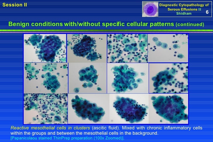
The mesothelial cells in this fluid resemble those seen in many non neoplastic effusions and essentially lack cytologic criteria of malignancy despite this fluid being from a dog with documented mesothelioma based on tissue invasion in surgical biopsies of tissues.
More From Sports Coloring Pages For Girls
- Columbia Mesothelioma Attorney
- Personal Injury Lawyer Staten Island
- Ip Law Firm Logo
- Crown Coloring Page Printable
- Doug Mesothelioma Commercials Still Alive
Incoming Search Terms:
- Pdf Cytopathologic Differential Diagnosis Of Malignant Mesothelioma Adenocarcinoma And Reactive Mesothelial Cells A Logistic Regression Analysis Funda Demirag Academia Edu Doug Mesothelioma Commercials Still Alive,
- The Don T Eat Me Signal Cd47 Is A Novel Diagnostic Biomarker And Potential Therapeutic Target For Diffuse Malignant Mesothelioma Abstract Europe Pmc Doug Mesothelioma Commercials Still Alive,
- Mesothelial Cytopathology Libre Pathology Doug Mesothelioma Commercials Still Alive,
- Integrative Approach To Cytologic And Molecular Diagnosis Of Malignant Pleural Mesothelioma Hjerpe Translational Lung Cancer Research Doug Mesothelioma Commercials Still Alive,
- What S New In Mesothelioma Pathologica Journal Of The Italian Society Of Anatomic Pathology And Diagnostic Cytopathology Doug Mesothelioma Commercials Still Alive,
- Effusion Cytology Diagnosis Doug Mesothelioma Commercials Still Alive,


