Pleural Thickening X Ray, What Is Pleural Thickening With Pictures
Pleural thickening x ray Indeed lately is being sought by users around us, perhaps one of you. Individuals now are accustomed to using the net in gadgets to view video and image information for inspiration, and according to the title of the article I will discuss about Pleural Thickening X Ray.
- Thorax 8 2 Pleural Space Case 8 2 2 Benign And Malignant Pleural Thickening Ultrasound Cases
- Tuberculous Pleural Effusion Shaw 2019 Respirology Wiley Online Library
- Clinical Diagnosis Of Malignant Pleural Mesothelioma Bianco Journal Of Thoracic Disease
- Pleura Chest Wall And Diaphragm Chest Radiology The Essentials 2nd Edition
- Epos Trade
- Preoperative Chest X Ray Showing Right Pleural Thickening With No Download Scientific Diagram
Find, Read, And Discover Pleural Thickening X Ray, Such Us:
- Pleural Plaques Causes Symptoms Diagnosis Treatment
- Personal Injury Claims The Difference Between Pleural Thickening And Pleural Plaques Tilly Bailey Irvine
- Epos Trade
- Case 20 1997 A 74 Year Old Man With Progressive Cough Dyspnea And Pleural Thickening Nejm
- Pleural Thickening And Pleural Calcification Radiology Key
- Occupations Causing Mesothelioma
- What Is Asbestos In Buildings
- Immigration Attorneys Mayra Joli
- Hartford Mesothelioma Lawyer
- Lol Pets Coloring Pages
If you are looking for Lol Pets Coloring Pages you've arrived at the ideal place. We have 104 graphics about lol pets coloring pages including images, photos, photographs, backgrounds, and more. In such web page, we additionally provide variety of graphics available. Such as png, jpg, animated gifs, pic art, symbol, black and white, transparent, etc.
Doctors can use a few different imaging scans to diagnose pleural thickening.

Lol pets coloring pages. Pleural plaques are patchy collections of hyalinized collagen in the parietal pleura. A ct scan which is also used to diagnose asbestosis and pleural plaques can confirm the condition earlier. Pet and mri scans provide even further detail and may be used to differentiate pleural thickening from malignant mesothelioma.
The most common way to diagnose pleural thickening is with imaging studies such as x rays mris or ct scans. It arises from a number of causes. If a patient is supine then a pleural effusion layers along the posterior aspect of the chest cavity and becomes difficult to see on a chest x ray.
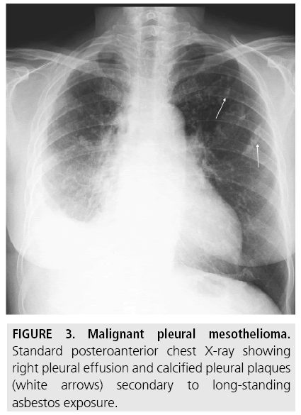
Pleural thickening leading to a malignant and benign lesion on chest x ray and ct scan has a diffe rent characteristic. An x ray can reveal thickening of the pleura but a ct scan is more likely to show thickening in better detail. Noncalcified pleural plaques are difficult to identify on the chest radiograph except when the x ray beam is tangential to the plaque.
Although pleural thickening is a common finding on routine chest x rays its radiological and clinical features remain poorly characterized. Our investigation of 28727 chest x rays obtained from annual health examinations confirmed that pleural thickening was the most common abnormal radiological finding. Right hilum is pulled upwards suggestive of volume loss in upper zone.
If the patient is upright when the x ray is taken then fluid will surround the lung base forming a meniscus a concave line obscuring the costophrenic angle and part or all of the hemidiaphragm. They have a holly leaf appearance on x ray. Apical pleural cap refers to a curved density at the lung apex seen on chest radiograph.
My x ray report is as follows. They are indicators of asbestos exposure and the most common asbestos induced lesion. It can occur with both benign and malignant pleural disease.
I have pleural thickening in left upper zone. 1 no other abnormality detected in lung fields. 3 both hila cpangles and diaphragm appear normal.
The plaque appears in profile as a sharply marginated dense band of soft tissue ranging from 1 to 10 mm in thickness paralleling the inner margin of the lateral thoracic wall. They usually appear after 20 years or more of exposure and never degenerate into mesothelioma. 2 heart and mediastinal markings appear normal.
Chronic ischemic etiology is favored for most cases 4. In most cases 922 pleural thickening involved the apex of the lung particularly on. The condition is usually first spotted through a chest x ray in which pleural thickening appears as an irregular shadow of the pleura.
According to etiology it may be classified as. Malignant mesothelioma malig nant pleural thickening has lesion characteristic of irregular nodular opacities that can be seen on. 4 bony thoracic case appears normal.
More From Lol Pets Coloring Pages
- Sarcomatoid Mesothelioma Life Expectancy
- What Does Asbestos Look Like In Walls
- T Rex Coloring Pages For Preschoolers
- Computer Coloring Pages
- Mesothelioma Financial Assistance
Incoming Search Terms:
- The Pleural Space Veterian Key Mesothelioma Financial Assistance,
- Apical Pleural Cap Radiology Reference Article Radiopaedia Org Mesothelioma Financial Assistance,
- Tuberculosis Radiology Wikipedia Mesothelioma Financial Assistance,
- Cxr Pneumothorax Pleural Thickening Mesothelioma Financial Assistance,
- Https Encrypted Tbn0 Gstatic Com Images Q Tbn 3aand9gcrvetogvgzcxcczyezo 07rlu6eymcoztt5wc3hudadudajfhv1 Usqp Cau Mesothelioma Financial Assistance,
- Http Pdf Posterng Netkey At Download Index Php Module Get Pdf By Id Poster Id 105668 Mesothelioma Financial Assistance,


