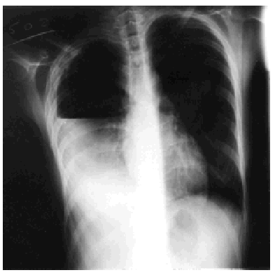
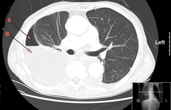
Pleural Thickening Chest X Ray, Pleural Thickening Causes Symptoms Treatment
Pleural thickening chest x ray Indeed recently has been hunted by users around us, perhaps one of you personally. Individuals now are accustomed to using the internet in gadgets to see video and image data for inspiration, and according to the name of this article I will discuss about Pleural Thickening Chest X Ray.
- Malignant Mesothelioma Presenting After Resolution Of A Seemingly Benign Asbestos Related Pleural Effusion European Medical Journal
- Early Detection Of Malignant Pleural Mesothelioma
- Pleural Thickening On Screening Chest X Rays A Single Institutional Study Respiratory Research Full Text
- Nodular Pleural Thickening In A Young Woman The Bmj
- Jaypeedigital Ebook Reader
- Dr Pepe S Diploma Casebook The Art Of Interpretation Case 134 Solved European Diploma Of Radiology
Find, Read, And Discover Pleural Thickening Chest X Ray, Such Us:
- Clinical Consequences Of Asbestos Related Diffuse Pleural Thickening A Review Journal Of Occupational Medicine And Toxicology Full Text
- Chest Radiograph Wikipedia
- Fibrothorax Wikiwand
- The Radiology Assistant Chest X Ray Basic Interpretation
- Tuberculous Pleural Effusion Shaw 2019 Respirology Wiley Online Library
- Lung Cell Structure
- Harry Potter Pumpkin Carving Patterns
- Mermaid Adult Coloring Book
- Free Online Coloring Pages
- Pd 1 Pd L1 And Mesothelioma
If you re searching for Pd 1 Pd L1 And Mesothelioma you've arrived at the ideal location. We have 104 graphics about pd 1 pd l1 and mesothelioma including pictures, photos, photographs, wallpapers, and much more. In such page, we also have variety of images available. Such as png, jpg, animated gifs, pic art, logo, black and white, transparent, etc.

Recurrent Tuberculous Pneumothorax And Tuberculous Empyema An Association Of Two Rare Complications Archivos De Bronconeumologia Pd 1 Pd L1 And Mesothelioma
Our investigation of 28727 chest x rays obtained from annual health examinations confirmed that pleural thickening was the most common abnormal radiological finding.

Pd 1 pd l1 and mesothelioma. Thickening of the pleural membranes is not a condition which is treatable. This should lead to immediate assessment of the patients trachea and mediastinum both on the x ray and more importantly clinically. Unilateral irregular nodular and diffuse pleural thickening is the classic finding on chest radiographs in patients with malignant mesothelioma.
Apical pleural cap refers to a curved density at the lung apex seen on chest radiograph. Pleural thickening is a common finding on routine chest x rays. 1012 the pleural thickening may be either plaquelike or nodular.
Fluid gathers in the lowest part of the chest according to the patients position. Chest pain and difficulty my xray report is as follows i have pleural thickening in left upper zone. If the lung edge visceral pleura is visible and there is black surrounding this edge then a pneumothorax should be suspected.
The chest xray is the most frequently requested radiologic examination. Chronic ischemic etiology is favored for most cases 4. What is pleural thickening.
Pleural thickening is a descriptive term given to describe any form of thickening involving either the parietal or visceral pleura. According to etiology it may be classified as. This pleural symptom can also occur in conjunction with a condition called hemothorax which is caused by the presence of blood in the chest cavity.
The frequency of apical pleural thickening increases with age 3. Publicationdate february 18 2013. Make sure you can see lung markings all the way to the edge of the chest wall.
A pleural effusion is a collection of fluid in the pleural space. Thickening of the pleura is a symptom of both asbestosis and mesothelioma but has other causes not related to asbestos. It typically involves the apex of the lung which is called pulmonary apical cap.
It arises from a number of causes. It can occur with both benign and malignant pleural disease. Tuberculosis caused by lung infection with mycobacterium tuberculosis and sarcoidosis a chronic inflammatory disease can also cause pleural thickening.
It typically involves the apex of the lung which is called pulmonary apical cap. If the patient is upright when the x ray is taken then fluid will surround the lung base forming a meniscus a concave line obscuring the costophrenic angle and part or all of the hemidiaphragm. However the fissures may also become.
Pleural thickening in chest x ray image results. Chest x ray screening pleural thickening pulmonary apical cap body mass index background pleural thickening is a common finding on routine chest x rays. On chest x rays the apical cap is an irregular density located at the extreme apex and is less than 5 mm in width 1.
Pleural effusions may obscure the pleura making it difficult to evaluate the thickness. On chest x rays the apical cap is an irregular density located at the extreme apex and.
More From Pd 1 Pd L1 And Mesothelioma
- Princess Coloring Online
- Peritoneal Mesothelioma Latency
- Mindfulness Colouring Sheets For Kids
- Fall Leaves Coloring Pages For Adults
- Vintage Pickup Truck Coloring Pages
Incoming Search Terms:
- Dr Pepe S Diploma Casebook The Art Of Interpretation Case 134 Solved European Diploma Of Radiology Vintage Pickup Truck Coloring Pages,
- Malignant Mesothelioma Presenting After Resolution Of A Seemingly Benign Asbestos Related Pleural Effusion European Medical Journal Vintage Pickup Truck Coloring Pages,
- Pleural Plaque Radiology Reference Article Radiopaedia Org Vintage Pickup Truck Coloring Pages,
- View Image Vintage Pickup Truck Coloring Pages,
- Thorax 8 2 Pleural Space Case 8 2 2 Benign And Malignant Pleural Thickening Ultrasound Cases Vintage Pickup Truck Coloring Pages,
- Pleural Thickening Costophrenic Angle Vintage Pickup Truck Coloring Pages,


