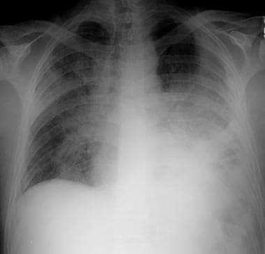
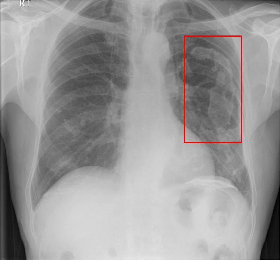
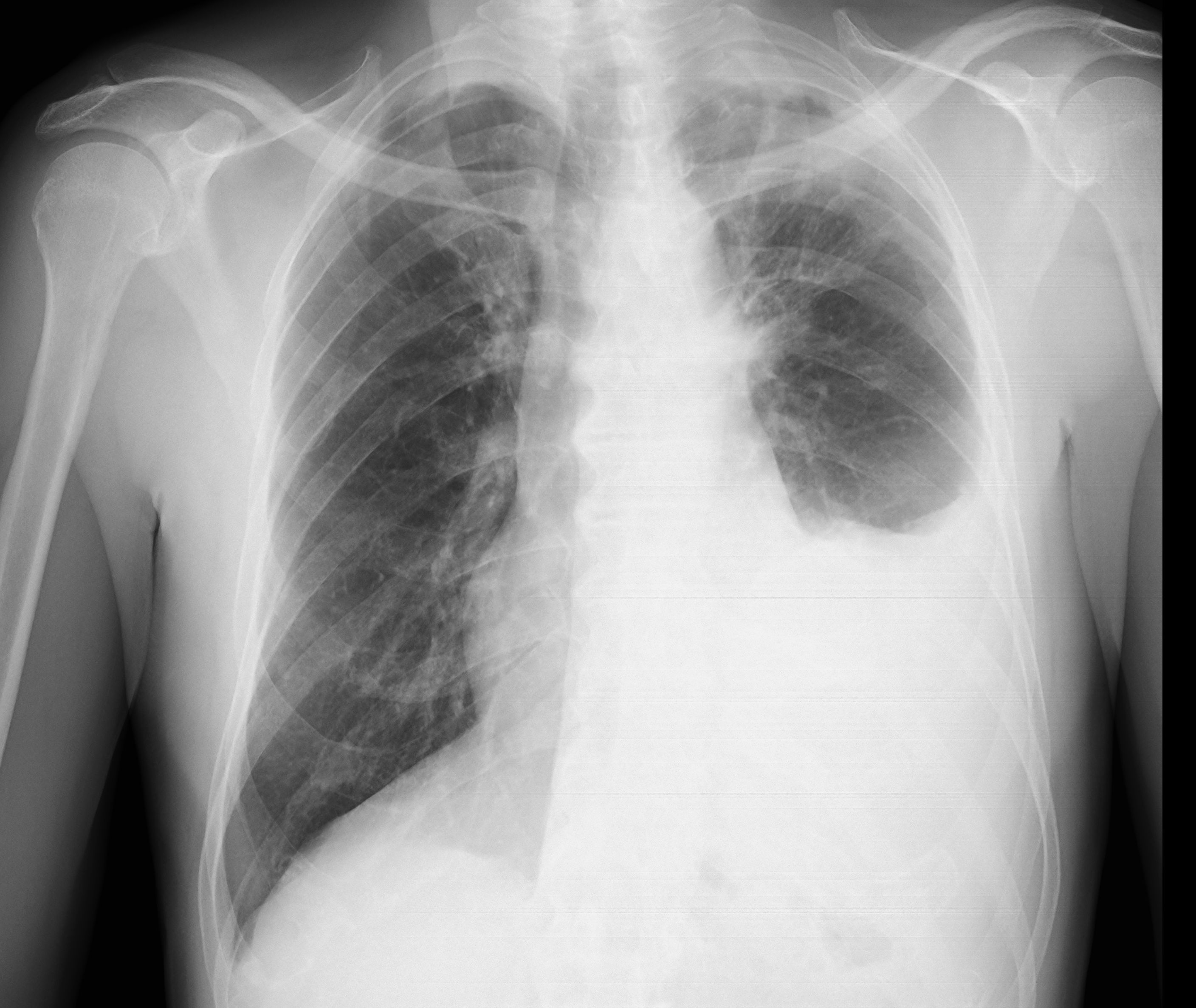
Pleural Thickening X Ray Images, Frontal Chest Radiograph Shows Bilateral Calcified Pleural Thickening There Is A Subtle Increase In Density Of The Central Inferi Radiology Radiographer Chest
Pleural thickening x ray images Indeed lately is being hunted by users around us, perhaps one of you personally. Individuals now are accustomed to using the net in gadgets to see video and image data for inspiration, and according to the name of this article I will talk about about Pleural Thickening X Ray Images.
- Learning Radiology Asbestos Related Pleural Disease Asbestosis
- Chest X Ray Showing Pleural Thickening Download Scientific Diagram
- Diagnostic Imaging And Workup Of Malignant Pleural Mesothelioma
- Asbestos When The Dust Settles An Imaging Review Of Asbestos Related Disease Radiographics
- Pleuroparenchymal Disease In A Ship Repair And Maintenance Worker
- Calcified Pleural Thickening
Find, Read, And Discover Pleural Thickening X Ray Images, Such Us:
- Benign Pleural Thickening Radiology Key
- Cxr Pneumothorax Pleural Thickening
- Apical Pleural Cap Radiology Reference Article Radiopaedia Org
- Chest Radiograph Shows Inhomogeneous Opacification Of L Open I
- Benign Diffuse Pleural Thickening Eurorad
- Christmas Mosaic Coloring Pages
- Warren Zevon Lawyers Guns And Money
- Painting Asbestos Ceiling Tiles
- Sharpie Coloring Pages
- Pictures Of Scary Carved Pumpkins
If you re searching for Pictures Of Scary Carved Pumpkins you've reached the right location. We have 104 graphics about pictures of scary carved pumpkins including images, photos, photographs, backgrounds, and much more. In these web page, we additionally have number of graphics out there. Such as png, jpg, animated gifs, pic art, logo, black and white, transparent, etc.
Chest x ray image of a healthy 47 year old male.
Pictures of scary carved pumpkins. However it should be noted that on a routine erect chest x ray as much as 250 600 ml of fluid is required before it becomes evident 6. Right hilum is pulled upwards suggestive of volume loss in upper zone. Hover onoff image to showhide findings.
If the asbestos degrades and is exposed asbestos fibres will be released into the. In most cases 922. Pleural thickening is best seen at the lung edges where the pleura runs tangentially to the x ray beam.
X ray of the chest of a 64 year old man with pleural thickening and calcification consistent with previous asbestos exposure. I have pleural thickening in left upper zone. Normal pleura and pleural spaces.
Pleural thickening is a descriptive term given to describe any form of thickening involving either the parietal or visceral pleura. Tutorial on chest x ray anatomy. However the fissures may also become.
Pleural effusions may obscure the pleura making it difficult to evaluate the thickness. Noncalcified pleural plaques are difficult to identify on the chest radiograph except when the x ray beam is tangential to the plaque. Unilateral irregular nodular and diffuse pleural thickening is the classic finding on chest radiographs in patients with malignant mesothelioma.
Other pleural diseases lead to fluid accumulation pleural effusion or air gathering in the pleural spaces pneumothorax. 1 no other abnormality detected in lung fields. The prevalence of pleural thickening was 32 n 91128727 in our sample.
The plaque appears in profile as a sharply marginated dense band of soft tissue ranging from 1 to 10 mm in thickness paralleling the inner margin of the lateral thoracic wall. Representative chest x ray image of pleural thickening. Pleural thickening due to asbestos.
3 both hila cpangles and diaphragm appear normal. My x ray report is as follows. Tap onoff image to showhide.
Unilateral pleural thickening hover onoff image to showhide findings. 1012 the pleural thickening may be either plaquelike or nodular. 2 heart and mediastinal markings appear normal.
Chest radiographs are the most commonly used examination to assess for the presence of a pleural effusion. Although pleural thickening is a common finding on routine chest x rays its radiological and clinical features remain poorly characterized. Our investigation of 28727 chest x rays obtained from annual health examinations confirmed that pleural thickening was the most common abnormal radiological finding.
A lateral decubitus projection is most sensitive able to identify even a small amount of fluid. The right apical cap appears as an irregular wedge shaped density. According to etiology it may be classified as.
It can occur with both benign and malignant pleural disease. 4 bony thoracic case appears normal.

Empyema Imaging Practice Essentials Radiography Computed Tomography Pictures Of Scary Carved Pumpkins
More From Pictures Of Scary Carved Pumpkins
- Ladybird Colouring Sheet
- Nixon Peabody Law Firm
- Hulk Colouring Sheets
- Immunohistochemical Diagnosis Of Mesothelioma
- Ropes And Gray Law Firm
Incoming Search Terms:
- Benign Pleural Thickening Radiology Key Ropes And Gray Law Firm,
- The Initial Chest X Ray Reveals A Pleural Thickening In The Right Lung Download Scientific Diagram Ropes And Gray Law Firm,
- Personal Injury Claims The Difference Between Pleural Thickening And Pleural Plaques Tilly Bailey Irvine Ropes And Gray Law Firm,
- Epos Ropes And Gray Law Firm,
- Radiological Review Of Pleural Tumors Sureka B Thukral Bb Mittal Mk Mittal A Sinha M Indian J Radiol Imaging Ropes And Gray Law Firm,
- Pleura Chest Wall And Diaphragm Chest Radiology The Essentials 2nd Edition Ropes And Gray Law Firm,


