Malignant Mesothelial Cells, Https Encrypted Tbn0 Gstatic Com Images Q Tbn 3aand9gcqipxph4wstcqoym53xfwxvcfrypiwahvqwk3iphwopz5jov0cm Usqp Cau
Malignant mesothelial cells Indeed lately is being sought by consumers around us, perhaps one of you. Individuals now are accustomed to using the internet in gadgets to see image and video data for inspiration, and according to the title of this post I will discuss about Malignant Mesothelial Cells.
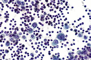
- Mesothelial Cells In Body Fluid Stock Photo Download Image Now Istock
- Benign Pleural Effusion With Ae1 Ae3 Positive Mesothelial Cells Mycytopathology
- View Image
- Positive Staining Of Mesothelial Cells Malignant Mesothelioma For Download Scientific Diagram
- 01 Presentation I Vs 8 55mb 3 28 08 Pps
- Http Www Asl5 Liguria It Portals 0 Anatomiapatologica2015 20150924 Effusion Cytology Pdf
Find, Read, And Discover Malignant Mesothelial Cells, Such Us:
- Https Onlinelibrary Wiley Com Doi Pdf 10 1002 Dc 20938
- Figure 1 From Cytological Diagnosis Of Malignant Mesothelioma Improvement By Additional Analysis Of Hyaluronic Acid In Pleural Effusions Semantic Scholar
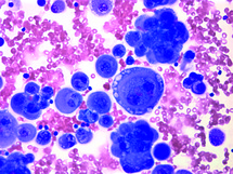
- Malignant Cells With Multiple Nuclei And Prominent Nucleoli Mesothelial Stock Photo Picture And Royalty Free Image Image 66564934
- Mesothelioma Malignant Mesothelioma The Cancer Of The Mesothelial Cells
- Reactive Mesothelial Hyperplasia Springerlink
- Mesothelioma Pleural Nodules
- Unicorn Cloud Coloring Pages
- Washington Mesothelioma Attorney
- Vermiculite Asbestos
- Mesothelioma And Lung Cancer In Asbestos
If you are searching for Mesothelioma And Lung Cancer In Asbestos you've reached the perfect place. We ve got 104 graphics about mesothelioma and lung cancer in asbestos including pictures, photos, photographs, backgrounds, and much more. In these webpage, we also provide number of graphics out there. Such as png, jpg, animated gifs, pic art, logo, black and white, transparent, etc.
The separation of benign from malignant mesothelial proliferations has emerged as a major problem in the pathology of the serosal membranes.

Mesothelioma and lung cancer in asbestos. Mesothelial cells are critical to the human body and are also involved in the transportation of fluids between body cavities and the transportation of cells for tissue repair or to combat inflammation. Benign mesothelial cells tend to arrange in monolayered sheets with little nuclear overlapping fig. The tumors can be either malignant cancerous or benign.
Mesothelial cells create a unique layer of paved like cells that line the bodys serious cavities and internal organs. To learn more about how cancers start and spread see what is cancer. These cells also cover the outer surface of most of your internal organs.
They contain ovoid nuclei fine chromatin delicate nuclear membrane small nucleoli and a moderate. This study tested the hypothesis that immunocytochemistry ic in effusion cell blocks cb can distinguish mm from rm and that the results may be applied to individual specimens. Malignant mesothelioma makes up the majority of diagnosed cases.
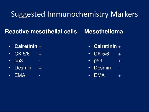
Distinguishing malignant mesothelioma mm from reactive mesothelial hyperplasia rm may be difficult in effusions. For tumor cells to take root and grow into masses in the mesothelium they must first find a way to infiltrate the membrane. The diagnosis of a mesothelial proliferation as malignant is most easily accomplished by identification of invasion of the mesothelial cells into underlying tissue lung skeletal muscle fibroadipose tissue etc and invasion can be highlighted with immunohistochemistry directed against cytokeratin andor calretinin.
For both epithelial and spindle cell mesothelial processes true stromal invasion is the most accurate indicator of malignancy but stromal invasion is often difficult to assess especially in small biopsies. Reactive mesothelial cells reactive mesothelial cells in pleural fluid reactive mesothelial cells are found when there is infection or inflammation present in a body cavity. The n body contains many different types of cells each of which has a specific function.
Neoplastic transformation of mesothelial cells results in malignant mesothelioma an aggressive tumor especially the pleura. Mesothelioma is categorized by the type of cells found in fluid or tissue samples taken from the body. The primary function of this layer termed the mesothelium is to provide a slippery non adhesive and protective surface.
A layer of specialized cells called mesothelial cells lines the inside of your chest your abdomen and the space around your heart.
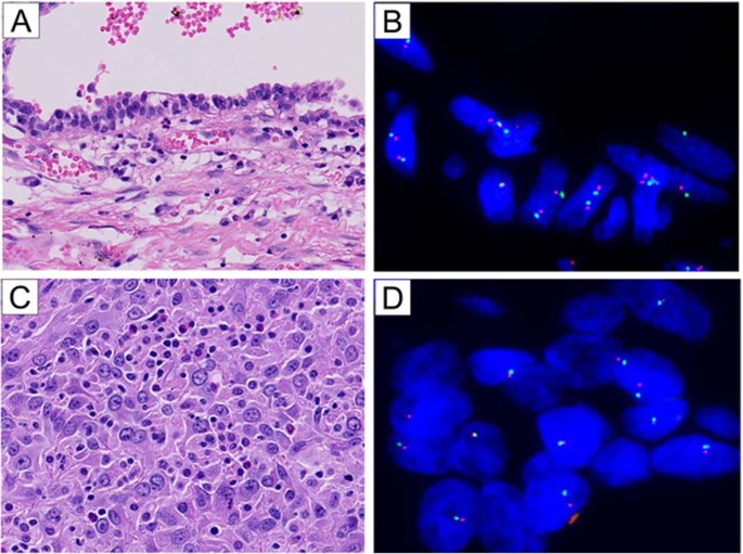
Hemizygous Loss Of Nf2 Detected By Fluorescence In Situ Hybridization Is Useful For The Diagnosis Of Malignant Pleural Mesothelioma Modern Pathology Mesothelioma And Lung Cancer In Asbestos
More From Mesothelioma And Lung Cancer In Asbestos
- Mesothelioma Resources
- Miner Mesothelioma
- Shopkins Coloring Pages Free Printable
- Printable Camel Pictures
- Creative Pumpkin Decorating Ideas
Incoming Search Terms:
- Combined Serum And Immunohistochemical Differentiation Between Reactive And Malignant Mesothelial Proliferations Sciencedirect Creative Pumpkin Decorating Ideas,
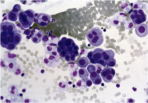
- Body Cavityfluids Chapter 3 Differential Diagnosis In Cytopathology Creative Pumpkin Decorating Ideas,
- Http Handouts Uscap Org 2016 Cm06 Daci 1 Pdf Creative Pumpkin Decorating Ideas,
- Utility Of Survivin Bap1 And Ki 67 Immunohistochemistry In Distinguishing Epithelioid Mesothelioma From Reactive Mesothelial Hyperplasia Creative Pumpkin Decorating Ideas,
- Pericardial Fluid Cytology A Large Irregular Cluster Of Malignant Download Scientific Diagram Creative Pumpkin Decorating Ideas,
- Mesothelial Cells In Body Fluid Stock Photo Download Image Now Istock Creative Pumpkin Decorating Ideas,
