Pleural Thickening Ultrasound, Https Adc Bmj Com Content Archdischild 104 1 12 Dc1 Embed Inline Supplementary Material 1 Pdf Download True
Pleural thickening ultrasound Indeed recently has been hunted by consumers around us, perhaps one of you. Individuals now are accustomed to using the internet in gadgets to view video and image information for inspiration, and according to the name of the post I will discuss about Pleural Thickening Ultrasound.
- Practice Pearls For Performing Pleural Ultrasound With Focus On Pleural Effusion And Pleural Thickening Touchrespiratory
- Wk 7 Non Cardiac Chest Thorax 8 2 Pleural Space Case 8 2 2 Benign And Malignant Pleural Thickening Ultrasound Cases Thorax Ultrasound Cardiac
- View Image
- Pleural Effusion Radiology Reference Article Radiopaedia Org
- Covid 19 Findings On Lung Ultrasound Thickened Pleural Line B Lines
- Sonographic Evaluation Of Pleural Effusion
Find, Read, And Discover Pleural Thickening Ultrasound, Such Us:
- Thorax 8 2 Pleural Space Case 8 2 2 Benign And Malignant Pleural Thickening Ultrasound Cases
- Emdocs Net Emergency Medicine Educationed Evaluation And Management Of Pleural Effusions One Size Doesn T Fit All Emdocs Net Emergency Medicine Education
- Perhimpunan Dokter Paru Indonesia
- Sonographic Evaluation Of Pleural Effusion
- Presentation1 Ultrasound Examination Of The Chest
- Rude Coloring Book
- Thing One Costume
- Mesothelioma Vs Squamous Cell Carcinoma
- Mesothelioma Asbestos Law Firm
- Meme Mesothelioma Button
If you are searching for Meme Mesothelioma Button you've come to the right place. We have 102 images about meme mesothelioma button including images, photos, photographs, backgrounds, and much more. In such webpage, we also provide number of graphics available. Such as png, jpg, animated gifs, pic art, symbol, black and white, translucent, etc.
Pleural thickening may occur in a variety of conditions.

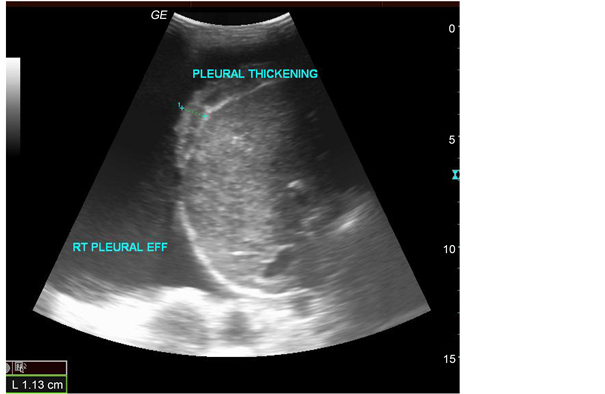
Meme mesothelioma button. It can occur from malignant as well as nonmalignant causes which include. Asbestos related pleural disease. Pleural thickening is often defined as a focal lesion that is greater than 3 mm in width arising from the visceral or parietal pleura with or without an irregular margin 13.
A suitable biopsy site is identified on ultrasound imaging and the depth from the skin to the parietal pleura is measured. It can occur with both benign and malignant pleural disease. As with pleural masses ultrasound or computed tomography ct may be essential for distinguishing loculated fluid.
Typically seen a continuous sheet of pleural thickening often involving the costophrenic angles and apices without calcification 2 3. Pleural effusion pleural thickening pleural. Using a tus threshold value of pleural thickening 1 cm as.
Chest ultrasound can supplement other imaging modalities of the chest and guides a variety of diagnostic and therapeutic procedures. Ultrasound has been proved to be valuable for the evaluation of a wide variety of chest diseases particularly when the pleural cavity is involved. When the pleural thickening is of recent onset days to weeks pleural effusion is the most likely cause of the opacity whereas if the process has been stable for months to years it is most probably true pleural thickening.
It is most frequently related to scarring fibrosis empyema and pleuritis. This procedure can be undertaken only if sufficient pleural fluid is present. Diffuse pleural fibrosis fibrothorax 6.
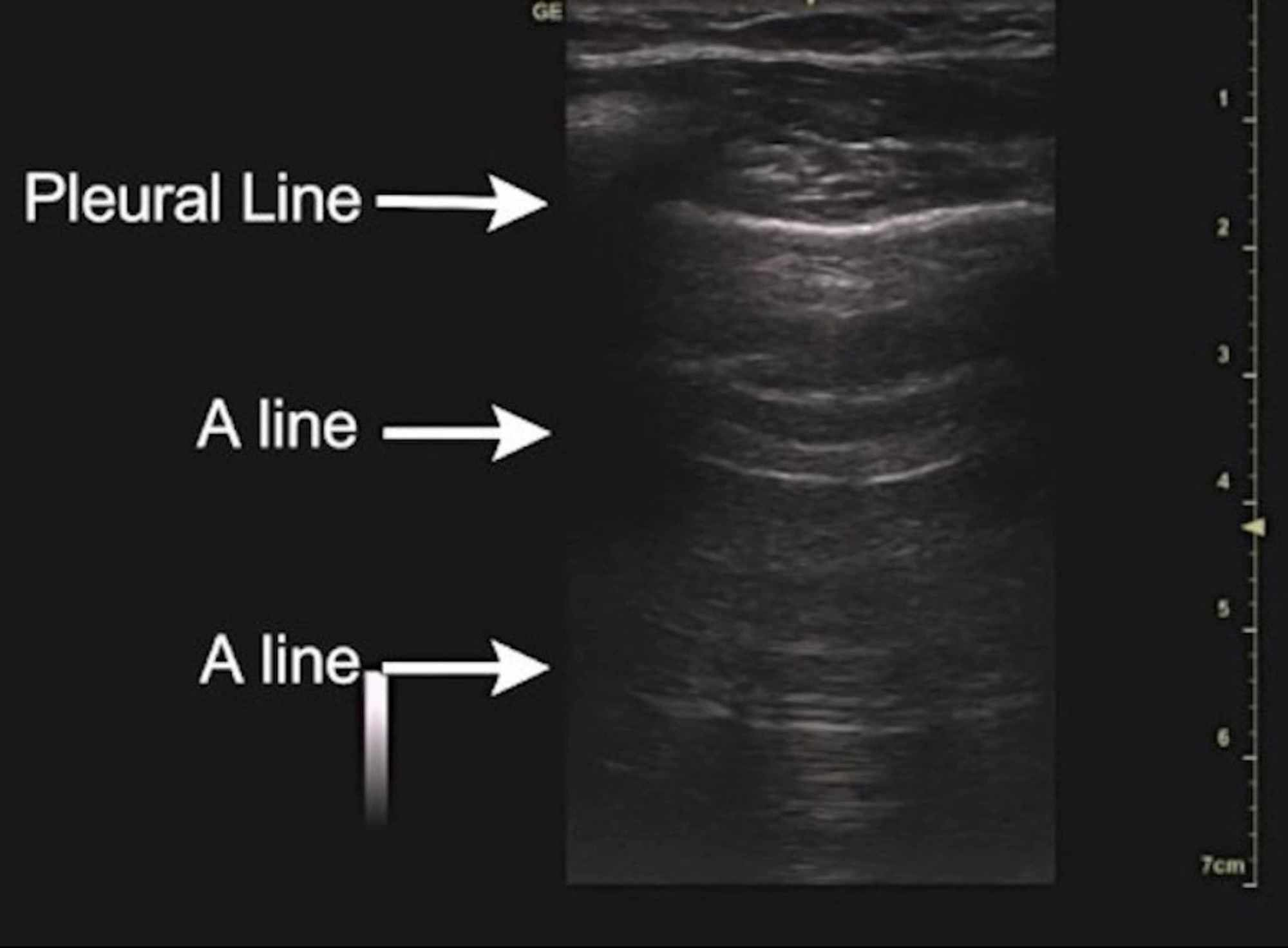
M mode identification of the pleural fluid sinusoid sign can also be helpful in distinguishing small effusions from pleural thickening. Unlike pleural effusion pleural thickening does not exhibit the fluid color sign movie 3. Diffuse pleural thickening refers to a morphological type of pleural thickening.
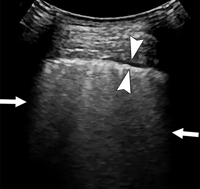
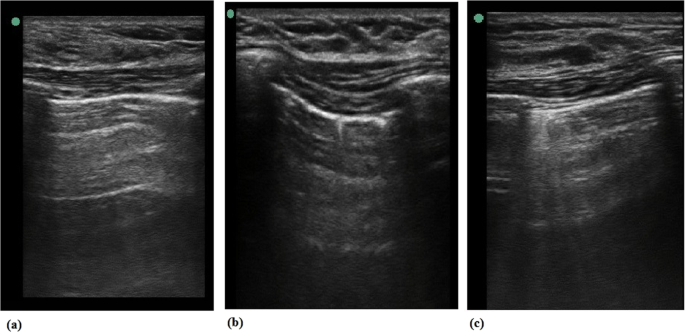
Pleural thickening appears as hypoechoic broadening of the pleura fig 4. 81 pulmonary pathology 82 pleural space 83 heart and mediastinum 84 thoracic wall. As most point of care pleural ultrasound in the intensive care unit is done to determine an optimal site for thoracentesis or tube thoracostomy making a fine distinction based on ultrasound between a very.
Parietal pleural thickening was detected in 21 patients measuring 1 cm in 1433 42 patients with a malignant effusion and 119 5 patients with a benign effusion x2 1df 811 p0004 and 1 cm in 233 6 malignant and 419 21 benign patients x2 1df 266 p010. Pleural thickening is a descriptive term given to describe any form of thickening involving either the parietal or visceral pleura. Distinguishing pleural thickening from small effusions can be challenging as both may appear hypoechoic on ultrasonography.
Https Adc Bmj Com Content Archdischild 104 1 12 Dc1 Embed Inline Supplementary Material 1 Pdf Download True Meme Mesothelioma Button
More From Meme Mesothelioma Button
- Jackson Law
- Mesothelioma And Cisplatin
- Fancy Bedroom Coloring Pages
- Zacchaeus Coloring Page
- Notifiable Asbestos
Incoming Search Terms:
- Presentation1 Ultrasound Examination Of The Chest Notifiable Asbestos,
- Dr Delgado Cidranes On Twitter Malignant Pleural Thickening The Rainbow Protocol The Art Of Lung Ultrasound Https T Co Mbtw2ybzac Foamed Notifiable Asbestos,
- Ultrasound Diagnosis Of Chest Diseaseses Intechopen Notifiable Asbestos,
- Assess Pulmonary Pathologies Using Vevo Ultrasound Fujifilm Visualsonics Notifiable Asbestos,
- Pleural Effusion Notifiable Asbestos,
- Ultrasound Of The Pleurae And Lungs Sciencedirect Notifiable Asbestos,


