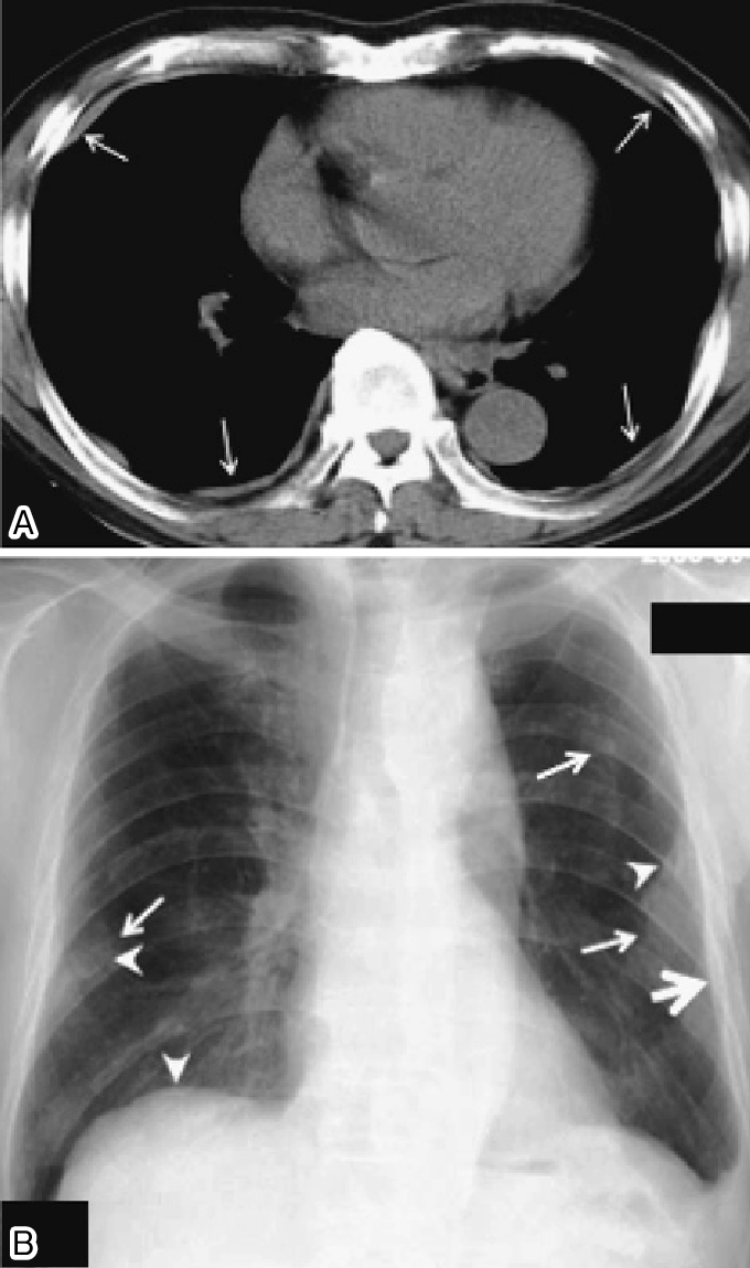
Pleural Plaques X Ray, Diagnostic Imaging And Workup Of Malignant Pleural Mesothelioma
Pleural plaques x ray Indeed lately is being sought by users around us, maybe one of you. People are now accustomed to using the internet in gadgets to see video and image information for inspiration, and according to the name of this article I will talk about about Pleural Plaques X Ray.
- Https Encrypted Tbn0 Gstatic Com Images Q Tbn 3aand9gcqfwdwi04cp9ib 2mgjcqq9larj1dru31gsd8pjk0v96i359xxr Usqp Cau
- Digital Chest Xray Showing Multiple Calcified Pleural Plaques From Asbestos Stock Photo Download Image Now Istock
- Loss Of Lung Markings Chest X Ray Medschool
- Pleural Thickening And Pleural Calcification Radiology Key
- Epos
- Pleural Changes Chest X Ray Medschool
Find, Read, And Discover Pleural Plaques X Ray, Such Us:
- Asbestos When The Dust Settles An Imaging Review Of Asbestos Related Disease Radiographics
- Pleural Plaques Asbestos Pa
- Pleural Plaques Holly Leaf Sign Radiology Radiology Student Medical Radiography
- Jaypeedigital Ebook Reader
- Asbestos Related Pleural Plaques Radiology Imaging
- Justice Law Firm
- Peritoneal Mesothelioma Cancer Types
- Thermal System Insulation Asbestos Type
- Choosing A Personal Injury Lawyer
- Cleaning Asbestos Floor Tiles
If you re searching for Cleaning Asbestos Floor Tiles you've come to the ideal location. We ve got 104 graphics about cleaning asbestos floor tiles including images, pictures, photos, wallpapers, and more. In such web page, we additionally provide variety of graphics available. Such as png, jpg, animated gifs, pic art, logo, black and white, transparent, etc.
Asbestos plaques example 3.

Cleaning asbestos floor tiles. Risk of mesothelioma this relates to the history of asbestos exposure and not to the presence of pleural plaques in general the finding of pleural plaques on a chest x ray in a patient with a history of asbestos exposure does not require formal follow up and the patient can be reassured 1. The diaphragm is often the best place to look for plaques where they lie in the plane of the x ray beam. Noncalcified pleural plaques are difficult to identify on the chest radiograph except when the x ray beam is tangential to the plaque.
Asbestos plaques example 3. Pleural plaques that do not calcify wont appear on an x ray and not all that do can be seen either it depends on the density of the calcification. Frontal multiple geographic areas of pleural calcification including involvement of the diaphragmatic surface.
When seen en face they may be difficult to see as is the left upper zone plaque in this image. Distinction of a loculated pleural effusion from pleural masses may not be possible on the chest x ray but should be easily distinguished on ct. Close up x ray of calcified asbestos pleural plaques.
The current study by wain as in the other studies does not carefully match asbestos exposure and smoking habits in those patients with pleural plaques vs those without. A pleural effusion is a collection of fluid in the pleural space. 2 case question available from the case.
Hover onoff image to showhide findings. Tap onoff image to showhide findings. Abstract pleural and subpleural pulmonary opacities may often be distinguished by their borders with tapered borders favoring a pleural origin while a sulcus sign or irregular borders favor a pulmonary origin.
The translucent white areas behind the rib cage show the pleural plaques. Plaque developing on lungs is considered an asbestos related disease but they are not cancerous. Pleural plaques exhibit the so called incomplete border sign on chest radiograph.
The inner margin is often well defined because it is tangential to the x ray beam and the adjacent lung is a good contrast medium. If the patient is upright when the x ray is taken then fluid will surround the lung base forming a meniscus a concave line obscuring the costophrenic angle and part or all of the hemidiaphragm. The tapering outer margin is indistinct as it is en face to the x ray beam and the chest wall provides less tissue contrast.
The plaque appears in profile as a sharply marginated dense band of soft tissue ranging from 1 to 10 mm in thickness paralleling the inner margin of the lateral thoracic wall. According to post mortem studies less than half of all cases of pleural plaque appear on an x ray. Pleural plaques in chest x rays of lung cancer patients and matched control.
This case demonstrates numerous calcified pleural plaques. Europ j respir dis.
More From Cleaning Asbestos Floor Tiles
- D Coloring Page
- Simpsons More Asbestos Gif
- Erin Brockovich Based On True Story
- Home Mesothelioma Treatment Asbestos
- Malignant Pleural Mesothelioma Review
Incoming Search Terms:
- What Is Pleural Plaques Amaa Malignant Pleural Mesothelioma Review,
- Calcified Pleural Plaques On Chest X Ray X Rays Case Studies Ctisus Ct Scanning Malignant Pleural Mesothelioma Review,
- Medpix Case Asbestos Related Pleural Plaques Malignant Pleural Mesothelioma Review,
- Pleural Calcifications Malignant Pleural Mesothelioma Review,
- Pulmonary Tuberculosis Complicating Asbestosis Hkmj Malignant Pleural Mesothelioma Review,
- Pleuroparenchymal Disease In A Ship Repair And Maintenance Worker Malignant Pleural Mesothelioma Review,


