Pleural Plaques Chest X Ray, Pleural Diseases Flashcards Memorang
Pleural plaques chest x ray Indeed recently is being sought by consumers around us, perhaps one of you personally. Individuals are now accustomed to using the net in gadgets to see video and image data for inspiration, and according to the title of this post I will discuss about Pleural Plaques Chest X Ray.
- Full Text Predictors Of Pneumonia On Routine Chest Radiographs In Patients With Copd
- Malignant Pleural Effusion Pulmonology Advisor
- Benign Pleural Thickening Radiology Key
- A Posteroanterior And B Lateral Chest Films Showed Extensive Download Scientific Diagram
- Asbestos Related Pulmonary Disorders Pulmonology Advisor
- View Image
Find, Read, And Discover Pleural Plaques Chest X Ray, Such Us:
- Pleural Plaque Radiology Reference Article Radiopaedia Org
- Pleuroparenchymal Disease In A Ship Repair And Maintenance Worker
- Digital Chest Xray Showing Multiple Calcified Pleural Plaques From Asbestos Stock Photo Download Image Now Istock
- Asbestos Related Pleural Plaques On Chest Xray Of Woman Stock Photo Download Image Now Istock
- Pulmonary Tuberculosis Complicating Asbestosis Hkmj
- Riddor Asbestos
- Mm Halloween Costume
- My Little Pony Printables Free
- Funny Cat Halloween Pictures
- Manufacturing Business And Mesothelioma
If you are searching for Manufacturing Business And Mesothelioma you've reached the ideal place. We ve got 100 graphics about manufacturing business and mesothelioma including pictures, photos, photographs, wallpapers, and more. In such webpage, we additionally have variety of images available. Such as png, jpg, animated gifs, pic art, symbol, blackandwhite, transparent, etc.
The diaphragm is often the best place to look for plaques where they lie in the plane of the x ray beam.

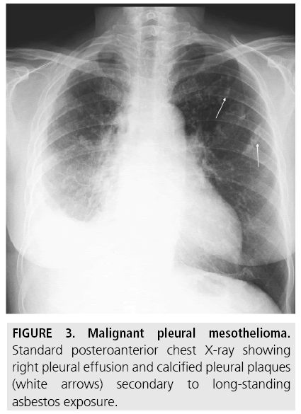
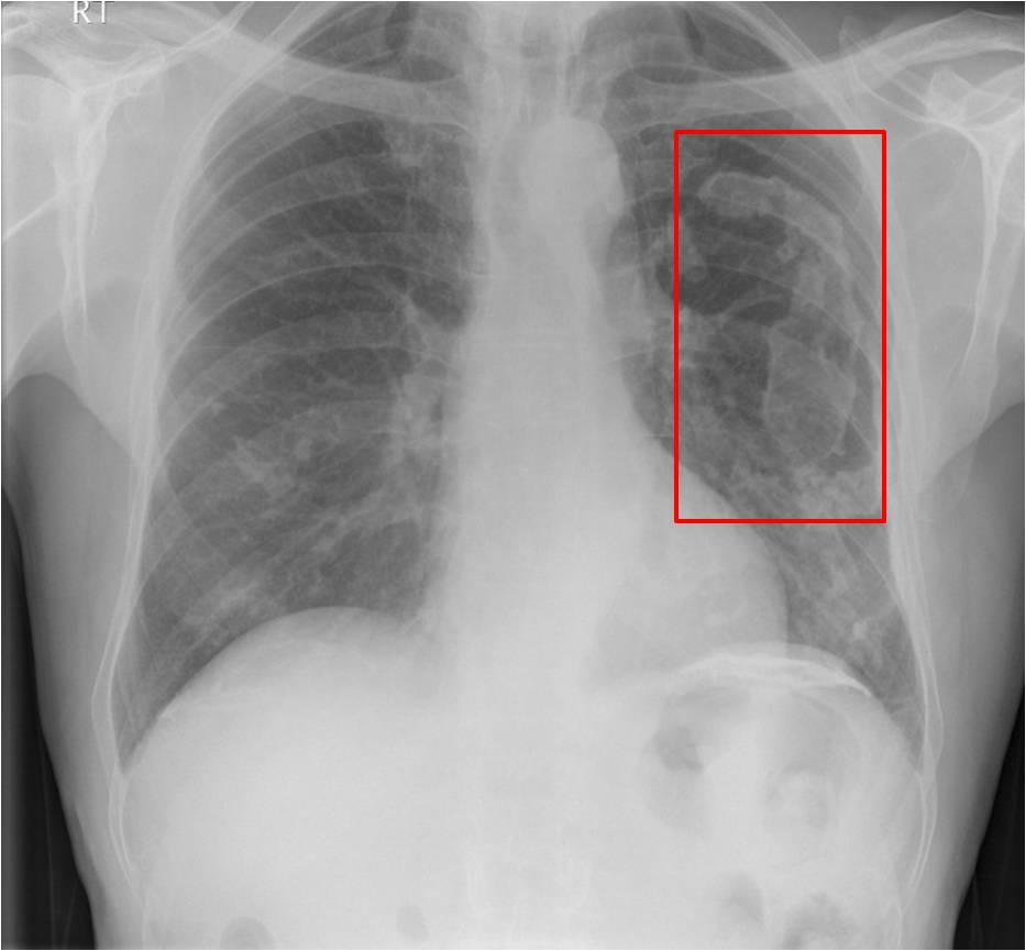
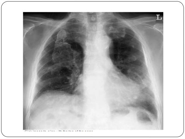
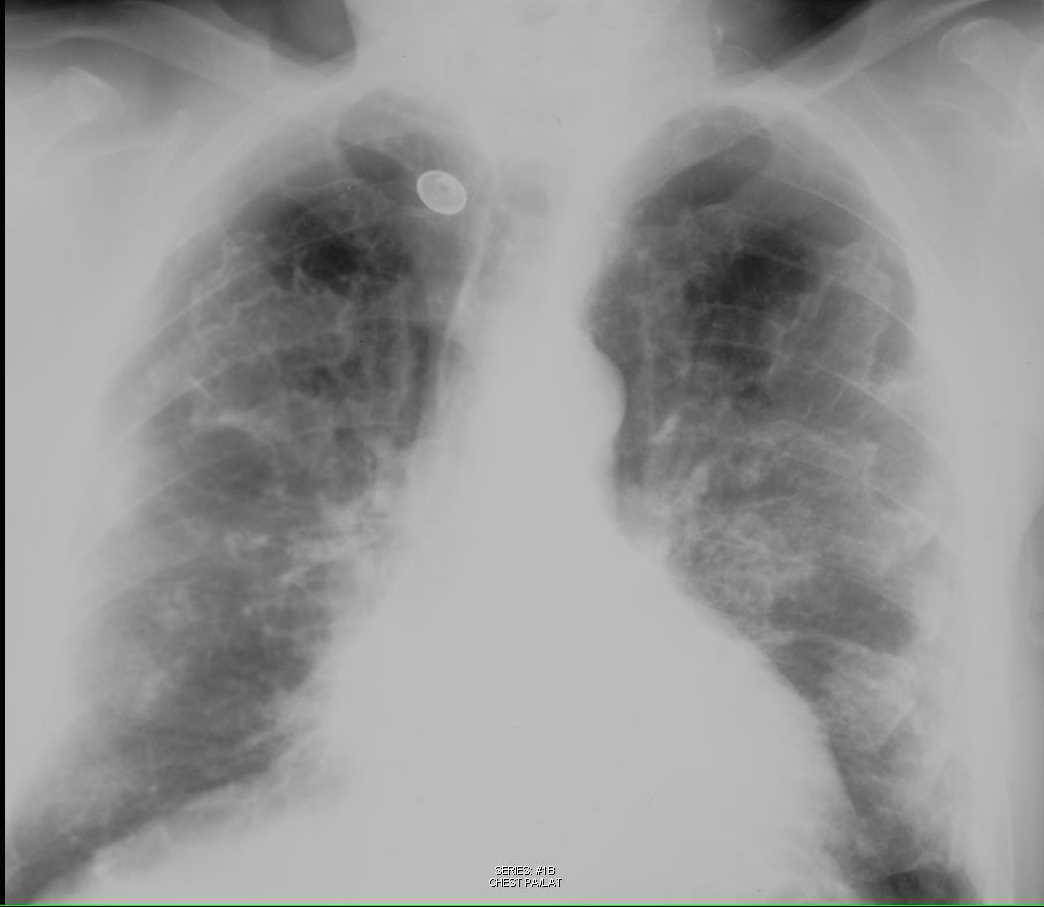
Manufacturing business and mesothelioma. Calcified pleural plaques appear as translucent white deposits on the lungs in x ray imaging scans. Asbestos plaques example 3. Abstract pleural and subpleural pulmonary opacities may often be distinguished by their borders with tapered borders favoring a pleural origin while a sulcus sign or irregular borders favor a pulmonary origin.
Adult chest x ray in the exam setting. A pleural effusion is a collection of fluid in the pleural space. Numbers and types of asbestos fibers in subjects with pleural plaques american journal of pathology november 1982 warnock et al.
Neonatal chest x ray in the exam setting. Diagnosis with ct scan a ct scan is the preferred method for diagnosing this condition as it can identify plaques anywhere in the chest even if they are not calcified. Commonly the condition may first be identified in a chest radiograph or x ray that shows thickened areas of the lung with concrete edges which some researchers have noted can look a bit like a holly leaf.
Pleural plaques are typically identified through imaging scans. Chest x ray in the exam setting. Risk of mesothelioma this relates to the history of asbestos exposure and not to the presence of pleural plaques in general the finding of pleural plaques on a chest x ray in a patient with a history of asbestos exposure does not require formal follow up and the patient can be reassured 1.
Their irregular thickened nodular edges are likened to the appearance of a holly leaf. Distinction of a loculated pleural effusion from pleural masses may not be possible on the chest x ray but should be easily distinguished on ct. Pediatric chest x ray in the exam setting.
If the patient is upright when the x ray is taken then fluid will surround the lung base forming a meniscus a concave line obscuring the costophrenic angle and part or all of the hemidiaphragm. When seen en face they may be difficult to see as is the left upper zone plaque in this image. The inner margin is often well defined because it is tangential to the x ray beam and the adjacent lung is a good contrast medium.
The tapering outer margin is indistinct as it is en face to the x ray beam and the chest wall provides less tissue contrast. Noncalcified pleural plaques frequently detected at autopsy are more difficult to appreciate radiographically as indicated by wain et al see page 707an increased sensitivity and awareness of pleural plaques and the use of oblique chest radiographs will increase the rate of detection. Pleural plaques exhibit the so called incomplete border sign on chest radiograph.
Asbestos plaques example 3. Tap onoff image to showhide findings. In all cases if your physician has seen evidence of pleural calcification on your chest x ray you should be checked for the presence of malignant cells in the area as well.
More From Manufacturing Business And Mesothelioma
- Cat In The Hat Coloring Pages To Print
- Dui Attorney Mesa Az
- Lol Dolls Colouring In Pictures
- Larsen Law
- Mesothelioma And
Incoming Search Terms:
- Chest X Rays 16 Subtle But Key Findings You Need To Know Mesothelioma And,
- Digital Chest Xray Of Asbestos Related Pleural Plaques And Mesothelioma Stock Photo Download Image Now Istock Mesothelioma And,
- Https Www Resmedjournal Com Article S0954 6111 17 30033 1 Pdf Mesothelioma And,
- 2 Radiology Of Pleural Disease Thoracic Key Mesothelioma And,
- Pleural Diseases Flashcards Memorang Mesothelioma And,
- Pleural Plaque Radiology Reference Article Radiopaedia Org Mesothelioma And,
