Pleural Mesothelioma Ct Images, Full Text Clinical Staging Of Malignant Pleural Mesothelioma Current Perspectiv Lctt
Pleural mesothelioma ct images Indeed recently is being hunted by consumers around us, maybe one of you. Individuals now are accustomed to using the internet in gadgets to view video and image information for inspiration, and according to the name of this article I will discuss about Pleural Mesothelioma Ct Images.
- Mesothelioma Radiology Reference Article Radiopaedia Org
- Volumetry An Alternative To Assess Therapy Response For Malignant Pleural Mesothelioma European Respiratory Society
- Malignant Pleural Mesothelioma Radiology Case Radiopaedia Org
- Mesothelioma Ct
- Mesothelioma Wikipedia
- Management Of Malignant Pleural Mesothelioma Part 1 Epidemiology Diagnosis And Staging Springerlink
Find, Read, And Discover Pleural Mesothelioma Ct Images, Such Us:
- Https Encrypted Tbn0 Gstatic Com Images Q Tbn 3aand9gcrk3iu5v9k5ajw7oamz6ty2mmk5ecwulxqpvc0sp37gjawyukbh Usqp Cau
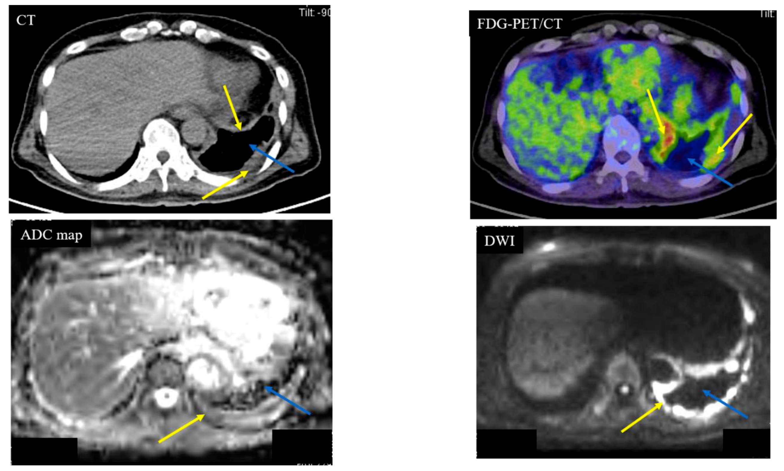
- Present And Future Roles Of Fdg Pet Ct Imaging In The Management Of Malignant Pleural Mesothelioma Semantic Scholar
- Management Of Malignant Pleural Mesothelioma Part 1 Epidemiology Diagnosis And Staging Springerlink
- Cureus Malignant Pleural Mesothelioma Biphasic Type An Unusual And Insidious Case Of Rapidly Progressive Small Blue Cell Tumor
- Low Dose Ct Screening May Increase Life Expectancy Of Pleural Mesothelioma Patients Mesowatch
- Mesothelioma Jl Steel
- Halloween Kids Colouring
- Pentecost Coloring Page
- Define Malignant Mesothelioma
- Printable Sunny Day Coloring Pages
If you are looking for Printable Sunny Day Coloring Pages you've arrived at the right location. We have 104 graphics about printable sunny day coloring pages adding pictures, pictures, photos, wallpapers, and much more. In these web page, we also have variety of graphics available. Such as png, jpg, animated gifs, pic art, symbol, black and white, translucent, etc.
Chaisaowong k aach t jaeger p et al.

Printable sunny day coloring pages. Ct scans enable doctors to identify the stage of a tumor by exposing. More is better a new report out of the uk suggests that a thorough diagnostic ct scan for pleural mesothelioma should include images of the abdomen and pelvis as well as the chest. Eighteen years experience in turkey.
Ct scans can help physicians to determine the stage of your tumor. Towards automated detection and quantitative assessment of pleural thickening from thoracic ct images. 12 clinical characteristics treatment and survival outcomes in malignant mesothelioma.
Still many doctors say the ct scan is the best for the chest and abdomen which are where mesothelioma forms. Diagnostic ct scan for mesothelioma. Use of ct for tumor staging.
Most tumors arise from the pleura and so this article will focus on pleural mesothelioma. Ct scans are preferred for staging tumors and are vital for patients with malignant pleural mesothelioma yendamuri explained. When a person goes to the doctor with early signs of malignant pleural mesothelioma a ct scan of the chest is often the next step in making a diagnosis.
Given the presence of the mesothelium in different parts of the body mesothelioma can arise in various locations 17. Computer assisted diagnosis for early stage pleural mesothelioma. Ct scans use x rays to capture detailed images from inside the body.
Differentials to consider would be pleural metastasis or invasive thymoma with pleural spread although these would be much less likely in this particular case. A new technique called ct perfusion can show if cancer cells are spreading in the bloodstream. The areas most prone to the formation of mesothelioma.
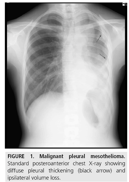
Malignant pleural mesothelioma mpm is the most common primary malignancy of the pleura and the second most common overall malignancy of the pleura after metastatic disease mpm arises from the mesothelial cells that cover the lung and chest wall and is strongly associated with asbestos exposure with latency periods ranging from 20 to 50 years. Ct scans are more than 90 sensitive for detecting malignant pleural mesothelioma. Appearances are typical of pleural mesothelioma especially given the evidence of underlying asbestos related pleural disease.
This scan also can show if it has spread to lymph nodes or other organs. Many radiologists favor the ct scan because of the great clarity in which the tumor image is represented. Although it is less effective at detecting peritoneal abdominal mesothelioma ct scans are still the most useful imaging study for diagnosing peritoneal mesothelioma.
Pleural mesothelioma 90 covered in this article. Pleural plaques are present in approximately 20 of mesothelioma patients.
More From Printable Sunny Day Coloring Pages
- Mesothelioma Paraneoplastic
- Easy Harry Potter Pumpkin Carving Patterns
- Elsa Printable Images
- Mesothelioma Vs Reactive Mesothelial Cells Cytology
- Mesothelioma Attorneys Navy Veteran
Incoming Search Terms:
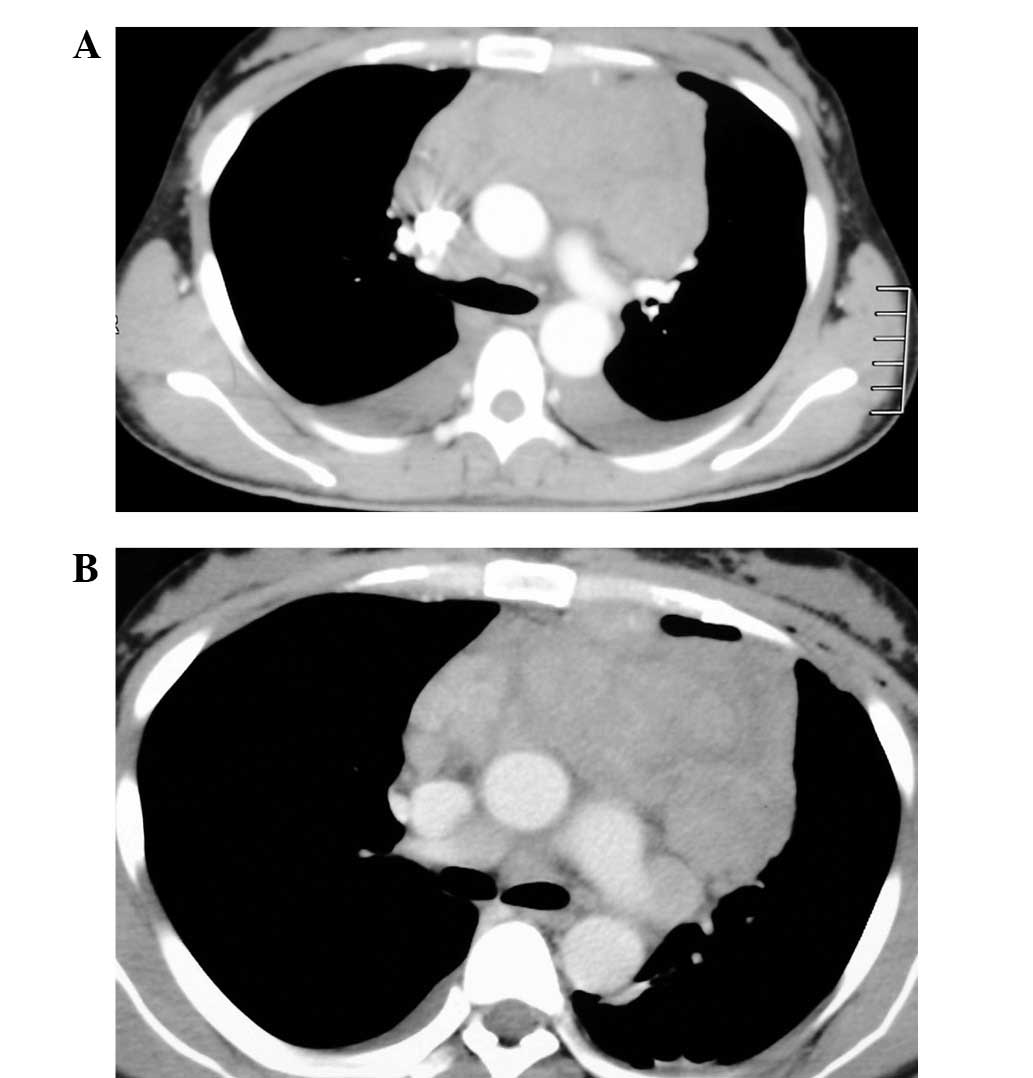
- A 71 Year Old Patient With Pleural Mesothelioma Ct Scan Top A Download Scientific Diagram Mesothelioma Attorneys Navy Veteran,
- Radiological Review Of Pleural Tumors Sureka B Thukral Bb Mittal Mk Mittal A Sinha M Indian J Radiol Imaging Mesothelioma Attorneys Navy Veteran,
- Malignant Mesothelioma Versus Metastatic Carcinoma Of The Pleura A Ct Challenge Iranian Journal Of Radiology Full Text Mesothelioma Attorneys Navy Veteran,
- Malignant Mesothelioma Versus Metastatic Carcinoma Of The Pleura A Ct Challenge Iranian Journal Of Radiology Full Text Mesothelioma Attorneys Navy Veteran,
- A Pseudo Mesothelioma Pleural Revelant An Carcinoma Urothelial Metastatic About A Case And Review Of The Literature Nnm Journal Of Lung Pulmonary Respiratory Research Mesothelioma Attorneys Navy Veteran,
- Mesothelioma Wikipedia Mesothelioma Attorneys Navy Veteran,


