Pleural Capping On Chest X Ray, Must Know Chest Radiograph Radiology Ppt Download
Pleural capping on chest x ray Indeed recently has been hunted by consumers around us, maybe one of you. Individuals now are accustomed to using the internet in gadgets to view image and video information for inspiration, and according to the name of this article I will discuss about Pleural Capping On Chest X Ray.
- Https Www Ajronline Org Doi Pdf 10 2214 Ajr 137 2 299
- Chest X Ray Note Left Pleural Effusion Pleural Thickening And Download Scientific Diagram
- Jaypeedigital Ebook Reader
- Pleura Chest Wall And Diaphragm Chest Radiology The Essentials 2nd Edition
- Case 20 1997 A 74 Year Old Man With Progressive Cough Dyspnea And Pleural Thickening Nejm
- Benign Pleural Thickening Radiology Key
Find, Read, And Discover Pleural Capping On Chest X Ray, Such Us:
- Apical Left Extrapleural Cap An Early And Important Sign On Chest Radiographs Emergency Medicine Journal
- Pleural Thickening Causes Symptoms Treatment
- Comparative Interpretation Of Ct And Standard Radiography Of The Pleura
- Pleura Chest Wall And Diaphragm Chest Radiology The Essentials 2nd Edition
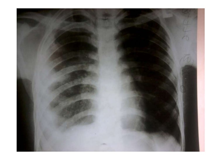
- B L Apices Collapsed With Rib Crowding Obvious Pleural Thickening B L Increased Vascularity Showing Pah Widening Of Heart Heart Shadow Radiology Thoracic
- Coloring Castle Shapes
- Mesothelioma Alimta Maintenance
- Symptoms Of Mesothelioma Prognosis
- Defense Attorney Public Defender Quotes
- Pearson Hardman Name Changes
If you re looking for Pearson Hardman Name Changes you've arrived at the right place. We have 104 graphics about pearson hardman name changes adding images, photos, photographs, wallpapers, and much more. In such webpage, we also have number of graphics out there. Such as png, jpg, animated gifs, pic art, symbol, black and white, translucent, etc.
It arises from a number of causes.

Pearson hardman name changes. Apical pleural cap radiology reference article. A pleural effusion is a collection of fluid in the pleural space. If the patient is upright when the x ray is taken then fluid will surround the lung base forming a meniscus a concave line obscuring the costophrenic angle and part or all of the hemidiaphragm.
It typically involves the apex of the lung which is called pulmonary apical cap. Chronic ischemic etiology is favored for most cases 4. Normal chest xray slideshare.
The frequency of apical pleural thickening increases with age 3. Five years previously she had had a normal chest roentgenogram fig 1 before needle drainage of bilateral axillary abscess. 1 svc 2 ivc 3 ra.
The pleural fluid can cap the apex of the lungs on a supine view fig. Fluid gathers in the lowest part of the chest according to the patients position. Pleural thickening is a common finding on routine chest x rays.
It typically involves the apex of the lung which is called pulmonary apical cap. On chest x rays the apical cap is an irregular density located at the extreme apex and is less than 5 mm in width. 137 of posts and discussions on chest x ray for pleural thickening.
Chronic ischaemic aetiology is favoured for most cases 4. Apical pleural cap refers to a curved density at the lung apex seen on chest radiograph. In the early twentieth cen tury a pulmonary apical cap was thought to be a.
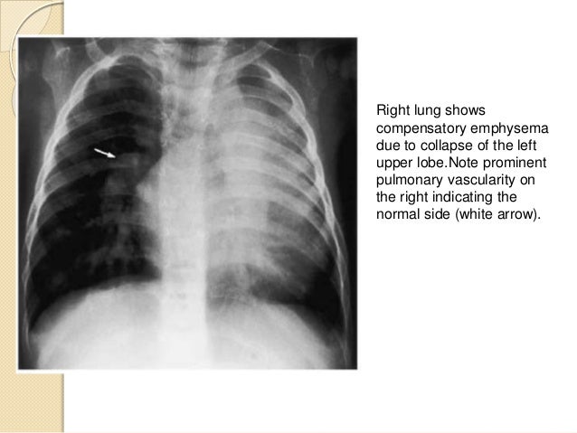
Increased homogeneous density superimposed over the lung loss of the hemidiaphragm silhouette blunted costophrenic angle apical capping elevation of the hemidiaphragm decreased visibility of lower lobe vasculature and accentuation of the minor fissure. On physical examination she had a continuous machinery like murmur heard predominantly on the second right intercostal space accompanied by a suprasternal systolic. It arises from a number of causes.
Apical pleural cap refers to a curved density at lung apex seen on chest radiographepidemiologythe frequency of apical pleural thickening apical pleural cap. Structures to be identified 3. 724 because of the relatively small capacity of the apex compared with the base of the thorax and because the superior and lateral aspects of the apex are the most dependent parts of the thorax tangential to a frontal x ray beam.
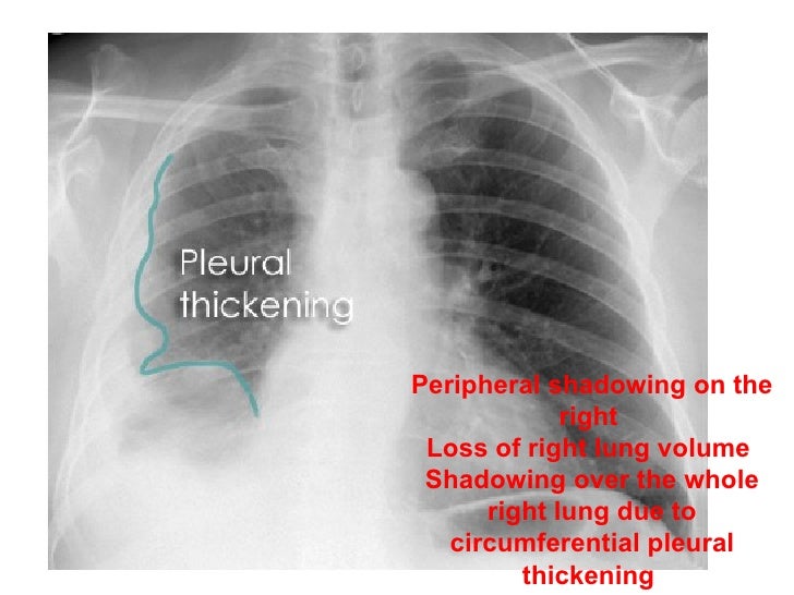
Pleural thickening is a descriptive term given to describe any form of thickening nodular pleural thickening. Pleural thickening and chest x ray treato. The frequency of apical pleural thickening increases with age 3.
A 20 year old asymptomatic woman was admitted to the hospital for workup of a continuous heart murmur. On chest x rays the apical cap is an irregular density located at the extreme apex and is less than 5mm in width 1. Apical pleural cap refers to a curved density at the lung apex seen on chest radiograph.
Normal chest xray 1. Pleural thickening is a common finding on routine chest x rays.
More From Pearson Hardman Name Changes
- What Is Mesothelioma Lawyer
- Twisty Petz Coloring Pages
- Well Differentiated Papillary Mesothelioma Treatment
- Cochran Law Firm
- Family Costume Ideas
Incoming Search Terms:
- Ct Chest Pleural Thickening Family Costume Ideas,
- Pleural Thickening Due To Asbestos X Ray Stock Image C038 2414 Science Photo Library Family Costume Ideas,
- Frontal Chest Radiograph Shows Bilateral Calcified Pleural Thickening There Is A Subtle Increase In Density Of The Central Inferi Radiology Radiographer Chest Family Costume Ideas,
- Case 20 1997 A 74 Year Old Man With Progressive Cough Dyspnea And Pleural Thickening Nejm Family Costume Ideas,
- Benign Pleural Thickening Radiology Key Family Costume Ideas,
- Chest Radiography Interpretation Ppt Video Online Download Family Costume Ideas,


