Peritoneal Fluid Mesothelial Cells Vs Macrophage, Http Www Cap Org Apps Docs Committees Hematology Educational Activities 2009 Cmb Pdf
Peritoneal fluid mesothelial cells vs macrophage Indeed lately is being hunted by consumers around us, perhaps one of you personally. People now are accustomed to using the net in gadgets to view video and image data for inspiration, and according to the title of the post I will discuss about Peritoneal Fluid Mesothelial Cells Vs Macrophage.
- Http Www Cap Org Apps Docs Committees Hematology Educational Activities 2009 Cmb Pdf
- Http Ar Iiarjournals Org Content 36 7 3579 Full Pdf
- Peritoneal Impact Of Il 17a Encyclopedia
- Mesothelial To Mesenchyme Transition As A Major Developmental And Pathological Player In Trunk Organs And Their Cavities Communications Biology
- A Cytocentrifugation Of Pleural Fluid Shows A Mixed Cell Population Download Scientific Diagram
- Cpd From Cloudy To Clear Effusions Made Easy Vet360
Find, Read, And Discover Peritoneal Fluid Mesothelial Cells Vs Macrophage, Such Us:
- Mesothelial Cell Csf1 Sustains Peritoneal Macrophage Proliferation Ivanov 2019 European Journal Of Immunology Wiley Online Library
- Http Ar Iiarjournals Org Content 36 7 3579 Full Pdf
- Hematology Cell Id Practical 1 Flashcards Quizlet
- Omentum Performs Unique Immunological Functions Medicine Sci News Com
- Cytological Diagnosis Of Peritoneal Endometriosis
- Free Printable Coloring Pages For Seniors
- Free Frida Kahlo Coloring Pages
- Mesothelioma Outcomes
- Mesothelioma Immunotherapy Australia
- Printable Masha Coloring Pages
If you re searching for Printable Masha Coloring Pages you've reached the ideal location. We ve got 104 images about printable masha coloring pages adding pictures, photos, pictures, backgrounds, and more. In such webpage, we also provide variety of images available. Such as png, jpg, animated gifs, pic art, symbol, blackandwhite, translucent, etc.
The proteins and serosal fluid trapped by the microvilli provide a slippery surface for internal organs to slide past one another.

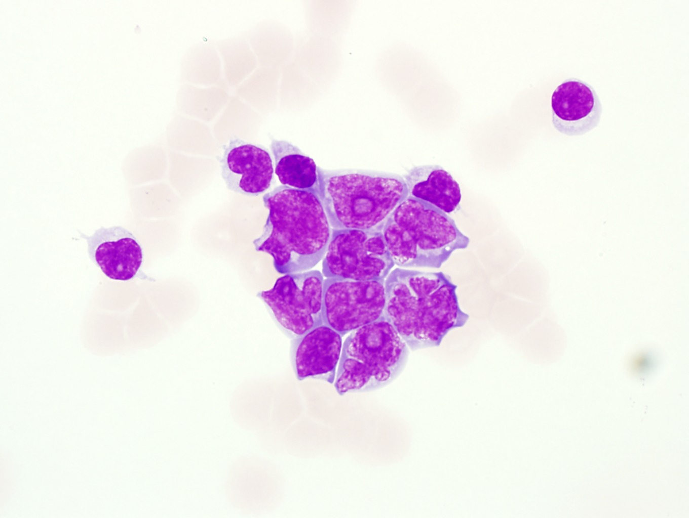
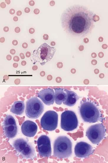
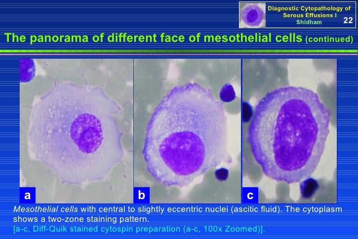
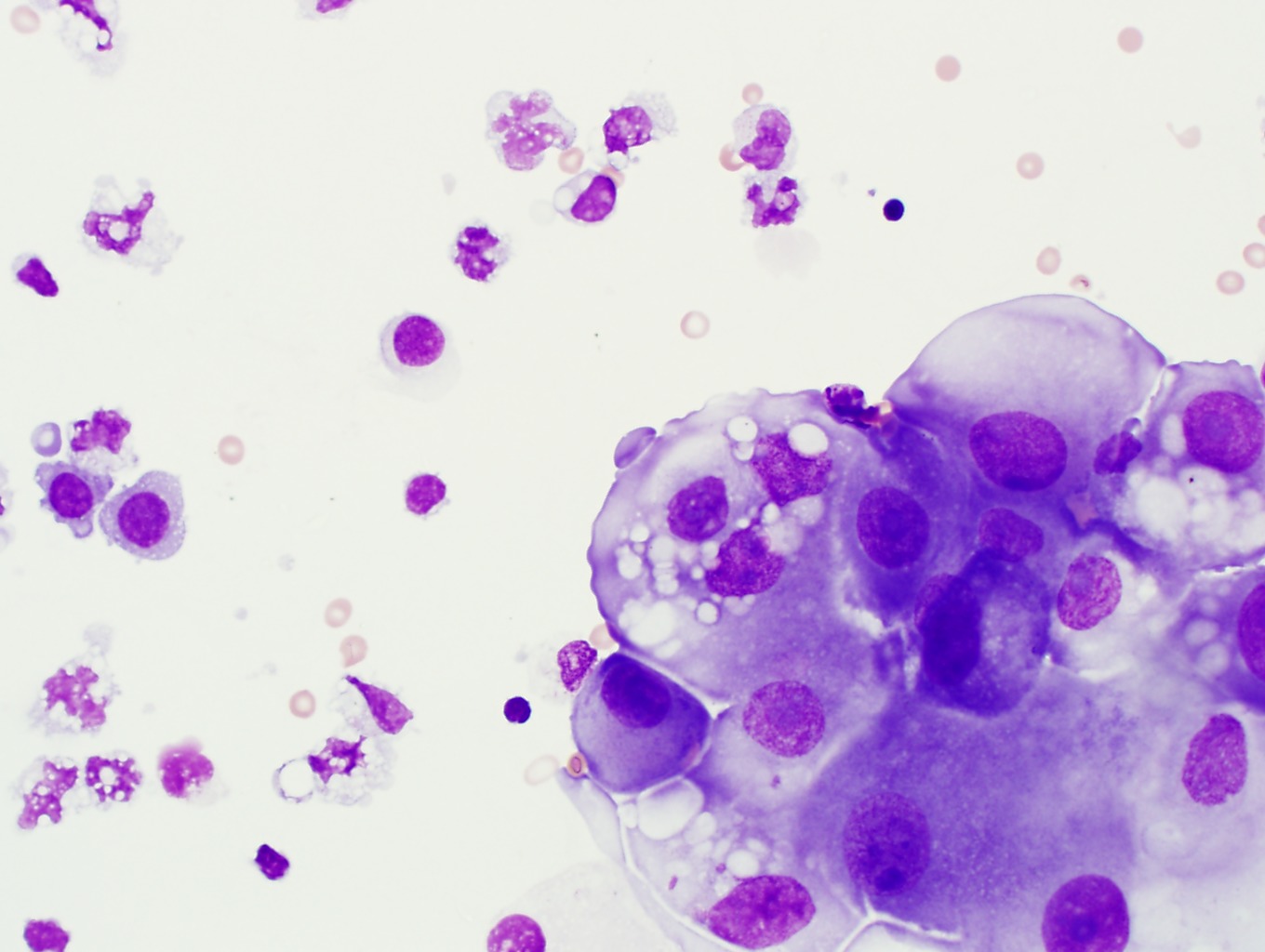
Printable masha coloring pages. Mesothelial cells have more abundant cytoplasm and rounder nuclei than monocytesmacrophages. Mesothelial cells are generally larger than monocytes and the cytoplasm does not normally contain phagocytized material. This course is intended for laboratory professionals who have experience with peripheral blood morphology and basic experience with body fluid differential analysisthis tutorial will provide a review of normal and abnormal body fluid morphology utilizing wright giemsa stained cytospin preparations from cerebrospinal fluid csf pleural peritoneal and synovial fluids as.
Cuboidal mesothelial cells may be found at areas of injury the milky spots of the omentum and the peritoneal side of the diaphragm overlaying the lymphatic lacunae. No significant differences have been identified between the mesothelial cells in the visceral or parietal pleura or among those in the pleural peritoneal or pericardial cavities. Variable but usually 5000ul unless there is blood contamination.
Red blood cell rbc count. J natl cancer inst. Uptake and transfer of particulate matter from the peritoneal cavity of the rat.
Microscopic examination of the pleural fluid revealed mostly neutrophils and occasional reactive and large multinucleated mesothelial cells demonstrating active. The amount of fluid is normally small less than 50 ml in humans and contains neutrophils mononuclear cells eosinophils macrophages lymphocytes desquamated mesothelial cells and an average of 30 gml of protein. Studies showed a wbc 2850ul and rbc 16800ul.
The arrowed cells are small lymphocytes one of which on right exhibits slight plasmacytoid features. Mixture of non degenerate neutrophils and macrophages roughly 5050 but can vary from 2080 to 8020 with low numbers of small lymphocytes and mesothelial cells. The nuclei should not be folded and in a reactive state they may have prominent nucleoli.
A phase contrast microscope study of free cells native to the peritoneal fluid of dba2 mice. The luminal surface is covered with microvilli. The two larger cells near the bottom with ruffled cytoplasmic.
Peritoneal fluid is produced by transudation from submesothelial vessels across the peritoneal membrane. Mesothelial cells can vary in shape from flat to cuboidal and in size ranging from approximately 10 to 50 mm in diameter and from 1 to over 4 mm in thickness. Research in recent years has examined the mechanisms underlying cellular host defence in the peritoneal cavity.
These studies have established that the resident cells of the peritoneal cavity the peritoneal macrophages pm phi and the mesothelial cells hpmc contribute to the initiation amplification and resolution of peritoneal inflammation. Mesothelial cell 38 01 this peritoneal fluid was obtained from a 74 year old man with congestive heart failure and ascites.
More From Printable Masha Coloring Pages
- Sun Coloring Pages Free
- Mesothelioma Cpc
- Coloring For Teens
- Pleural Metastasis
- Can Mesothelioma Be Caused By Rust
Incoming Search Terms:
- Https Www Rcpath Org Asset Ed8cdd8d 8d04 4b82 Ad48d585e2f023be Can Mesothelioma Be Caused By Rust,
- 9 Body Fluids Can Mesothelioma Be Caused By Rust,
- Cd90 Mesothelial Like Cells In Peritoneal Fluid Promote Peritoneal Metastasis By Forming A Tumor Permissive Microenvironment Can Mesothelioma Be Caused By Rust,
- Cancers Free Full Text With Great Age Comes Great Metastatic Ability Ovarian Cancer And The Appeal Of The Aging Peritoneal Microenvironment Html Can Mesothelioma Be Caused By Rust,
- Cytology Of Body Fluid Pleural Peritoneal Pericardial Ppt Video Online Download Can Mesothelioma Be Caused By Rust,
- Https S3 Amazonaws Com Ascpcdn Static Ascpresources Press Store Pdfs Body Fluid Analysis Lookinside 5b1 5d Pdf Can Mesothelioma Be Caused By Rust,
