Peripheral Lung Location, Jaypeedigital Ebook Reader
Peripheral lung location Indeed recently is being sought by consumers around us, maybe one of you personally. Individuals are now accustomed to using the net in gadgets to view image and video data for inspiration, and according to the title of the article I will talk about about Peripheral Lung Location.
- Lung Cancer Wikipedia
- Evaluation Of Various Cytological Examinations By Bronchoscopy In The Diagnosis Of Peripheral Lung Cancer British Journal Of Cancer
- Jaypeedigital Ebook Reader
- Different Histological Subtypes Of Peripheral Lung Cancer Based On Emphysema Distribution In Patients With Both Airflow Limitation And Ct Determined Emphysema Lung Cancer
- Schema Of Central And Peripheral Locations This Diagram Showed Download Scientific Diagram
- Lung Anatomy Physiopedia
Find, Read, And Discover Peripheral Lung Location, Such Us:
- Molecular Genetics Of Lung Cancer In People Who Have Never Smoked Sciencedirect
- Lung Auscultation Physical Therapy Reviewer
- Table 2 From Blind Spot In Lung Cancer Lymph Node Metastasis Cross Lobe Peripheral Lymph Node Metastasis In Early Stage Patients Semantic Scholar
- Jaypeedigital Ebook Reader
- Endobronchial Ultrasound Driven Biopsy In The Diagnosis Of Peripheral Lung Lesions Chest
- Free Printable Coloring Sheets For Kindergarten
- Happy Valentines Day Hearts Coloring Pages
- Mesothelioma Tumour Markers
- Disposable Asbestos Suits
- Mesothelioma Ct Scans
If you are searching for Mesothelioma Ct Scans you've come to the perfect place. We ve got 104 images about mesothelioma ct scans including images, photos, photographs, wallpapers, and much more. In such web page, we also provide variety of images available. Such as png, jpg, animated gifs, pic art, symbol, blackandwhite, transparent, etc.
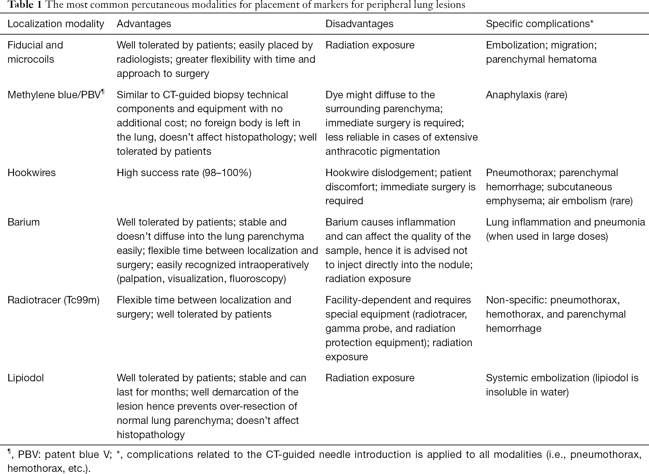
Placement Of Markers To Assist Minimally Invasive Resection Of Peripheral Lung Lesions Velasquez Annals Of Translational Medicine Mesothelioma Ct Scans

Different Histological Subtypes Of Peripheral Lung Cancer Based On Emphysema Distribution In Patients With Both Airflow Limitation And Ct Determined Emphysema Lung Cancer Mesothelioma Ct Scans
Within peripheral lung consolidations different entities show characteristic distribution patterns of pulmonary artery and bronchial artery flow signals in respect of frequency and location.
Mesothelioma ct scans. 34 b may be needed to confirm the peripheral location. A pulmonary embolism is also characterized as central or peripheral depending on the location or the arterial branch involved. 32 a or even a computed tomography ct scan 124 see fig.
Recognize a pattern of peripheral lung disease on chest radiography or computed tomography ct and give an appropriate differential diagnosis including a single most likely diagnosis when supported by associated radiologic findings or clinical information eg peripheral lung disease associated with paratracheal and bilateral hilar lymphadenopathy in an asymptomatic. Central tumors according to american college of chest physician accp guidelines are sampled easily under direct bronchoscopic visualization with an 88 percent diagnostic yield. Mnemonics for peripheral lung opacities seen on chest x ray or ct are useful to remember differentials.
A practical approach to evidence based clinical evaluation and management 2018. Ct guidance was performed by using single section ct equipment prospeed. 1 chest wall 2 pleura and 3 subpleural area of the lung.
I dont know about kosher but halal meat just requires the animal to die in the least amount of pain which at the the time the quran was written was to slit the. They are considered less common than the more centrally located bronchial carcinoid tumors. Reliable demarcation of vessels of tumor neo angiogenesis is currently not possible with color doppler.
Diagnostic yields using traditional methods are low 30 for peripheral lung nodules and are modest overall because before the development of ultrasound and navigation guided techniques biopsy was performed under. Nair md ms michael k. Aeiou sic cue mnemonics aeiou a.
Ge medical systems milwaukee wis from 2000 until 2004 and by using a 32 section multidetector ct system light speed xt. Lung tumors depending on their location in the tracheobronchial tree are categorized as central or peripheral. Gould md ms in lung cancer.
Peripheral pulmonary carcinoid tumor refer to a subtype of pulmonary carcinoid tumors that arise within the periphery of the lung. 32 b and fig. The lateral view see fig.
Arterial spectral curve analysis is time consuming.

Cancers Free Full Text Safety And Usefulness Of Cryobiopsy And Stamp Cytology For The Diagnosis Of Peripheral Pulmonary Lesions Html Mesothelioma Ct Scans
More From Mesothelioma Ct Scans
- Disney Coloring World
- Lawrence Parsons Lord Oxmantown
- Circulatory System Heart Coloring Sheet
- Commercial Real Estate Attorney
- Forest Coloring Pages For Adults
Incoming Search Terms:
- Early Diagnosis Of Solitary Pulmonary Nodules Xu Journal Of Thoracic Disease Forest Coloring Pages For Adults,
- Https Encrypted Tbn0 Gstatic Com Images Q Tbn 3aand9gcs Wddf Awcpnmjop8n12l 4omm5btl7 Dvbwx 9b7cqq2 Mk4 Usqp Cau Forest Coloring Pages For Adults,
- Https Encrypted Tbn0 Gstatic Com Images Q Tbn 3aand9gcquzbquqv Ugexsl5moemhzqviuckv Q Tzgfco7iiqdqvdxtrb Usqp Cau Forest Coloring Pages For Adults,
- Cancers Free Full Text Safety And Usefulness Of Cryobiopsy And Stamp Cytology For The Diagnosis Of Peripheral Pulmonary Lesions Html Forest Coloring Pages For Adults,
- Peripheral Pulmonary Stenosis Symptoms Causes In Children Boston Children S Hospital Forest Coloring Pages For Adults,
- Pdf Percutaneously Implanted Markers In Peripheral Lung Tumours Report Of Complications Lena Specht And Mirjana Josipovic Academia Edu Forest Coloring Pages For Adults,

