Mesothelioma Vs Reactive Mesothelial Cells, Common Immunohistochemical Stains Used To Differentiate Pulmonary Download Table
Mesothelioma vs reactive mesothelial cells Indeed lately is being sought by users around us, perhaps one of you. Individuals are now accustomed to using the internet in gadgets to view video and image data for inspiration, and according to the name of the article I will discuss about Mesothelioma Vs Reactive Mesothelial Cells.
- Pdf Epithelial Membrane Antigen In Differential Diagnosis Of Malignant Mesothelioma Metastatic Adenocarcinoma And Reactive Mesothelial Hyperplasia Malign Mezotelyoma Metastatik Adenokarsinoma Ve Reaktif Mezotelyal Hiperplazi Ayirici Tanisinda
- Http Handouts Uscap Org 2016 Cm06 Daci 1 Pdf
- Https Encrypted Tbn0 Gstatic Com Images Q Tbn 3aand9gctavdn5rq6ogml6mbrkvulkbg8b Tuympbyippf068sohuie1ny Usqp Cau
- Https Www Surgpath Theclinics Com Article S1875 9181 10 00011 5 Pdf
- Mesothelioma Dog
- Malignant Mesothelioma In Situ Morphologic Features And Clinical Outcome Modern Pathology
Find, Read, And Discover Mesothelioma Vs Reactive Mesothelial Cells, Such Us:
- Normal Cytology Springerlink
- Malignant Mesothelioma In Situ Morphologic Features And Clinical Outcome Modern Pathology
- Https Academic Oup Com Ajcp Article Pdf 110 3 397 24884795 Ajcpath110 0397 Pdf
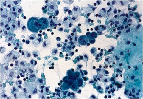
- Sheet Of Reactive Mesothelial Cells Note Owindowso Between Cells And Download Scientific Diagram
- Pathologic Diagnosis And Classification Of Mesothelioma Springerlink
- Halloween Party Photos
- Halloween A4 Colouring Pages
- Springtrap Coloring Pages
- Mesothelioma What Feelings Happens To You
- Mesothelioma Doctors In South Carolina
If you are looking for Mesothelioma Doctors In South Carolina you've reached the right location. We have 104 images about mesothelioma doctors in south carolina adding images, photos, pictures, wallpapers, and more. In such web page, we also have number of images out there. Such as png, jpg, animated gifs, pic art, symbol, blackandwhite, transparent, etc.
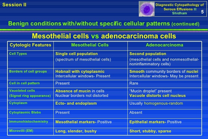
Cytologic differential prognosis amongst reactive.

Mesothelioma doctors in south carolina. Reactive mesothelial cells show no immunoreactivity. The condition can be due to bacterial viral or fungal infections. Immunohistochemical detection of glut 1 can discriminate between reactive mesothelium and malignant mesothelioma.
Likewise strong membranous positivity for glut 1 andor strong nuclear staining for p53 favors a mesothelioma. The reactive mesothelial hyperplasia can be so florid that it may histologically mimic even for an experienced pathologist some malignant tumors such as mesothelioma or metastasis of an. Mm 2 or more of the nucleus in an atypical in situ mesothelial lesion of the pleura are found consistently in neoplastic mesothelial cells.
Mesothelioma vs reactive mesothelial cells. Shilkin frcpath frcpa darrel whitaker phd reactive mesothelial hyperplasia vs mesothelioma including mesothelioma in situ. The differential diagnosis of epithelial type mesothelioma from adenocarcinoma and reactive mesothelial proliferation.
Reactive mesothelial cells are found when there is an infection in the body. Reactive mesothelial the evidence of underlying tissue infiltration by means of mesothelial cells presently remains the gold fashionable for. Conversely a combination of negative ema and positive desmin favors a reactive process.
Unique article cytologic differential analysis among reactive mesothelial cells malignant mesothelioma and adenocarcinoma. It also can be because of a tumor such as from mesothelioma. Initial workup could use 2 mesothelial markers and 2 markers for the other tumor in the differential diagnosis on the basis of morphology use more markers if results inconclusive imig international mesothelioma interest group recommends 2 mesothelial markers and 2 carcinoma markers be included in the panel.
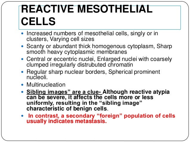
The reaction to injury determines that the mesothelial proliferation may exceed the normal regeneration resulting in a reactive mesothelial hyperplasia. Reactive mesothelial cells show strong membranous positivity and cytoplasmic staining. Malignant mesothelial cells show no to weak and focal staining.
E epithelial membrane antigen ema. Mesotelioma pleural maligno scielo espana. Comparison of 22 cases of mesothelioma in situ that fulfill these requirements for diagnosis with 141 invasive mesotheliomas and 78 reactive mesothelioses indicates that strong linear membrane related labeling for epithelial membrane antigen and silver labeled nucleolar organizer region positive material that occupies 06677 microm2 or more of the nucleus in an atypical in situ mesothelial lesion of the pleura are found consistently in neoplastic mesothelial cells.
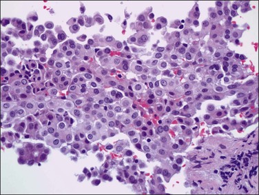
B an example of a cell block from a case of malignant mesothelioma is shown h e. These cells usually come in clumps and have more of a washed out cytoplasm in the bodily fluids. Reactive mesothelial the proof of underlying tissue infiltration by using mesothelial cells presently stays the gold trendy for.

Use Of Panel Of Markers In Serous Effusion To Distinguish Reactive Mesothelial Cells From Adenocarcinoma Subbarayan D Bhattacharya J Rani P Khuraijam B Jain S J Cytol Mesothelioma Doctors In South Carolina
More From Mesothelioma Doctors In South Carolina
- Treatment Of Benign Multicystic Peritoneal Mesothelioma
- First Female Lawyer Supreme Court In India
- Wonder Woman Printable Coloring Pages
- Rocky Horror Dress Up Ideas
- Sonic Printable Coloring Pages
Incoming Search Terms:
- Mesothelial Cytopathology Libre Pathology Sonic Printable Coloring Pages,
- Pleural Fluid All Cell Blocks A D Pleural Mesothelioma Epithelial Download Scientific Diagram Sonic Printable Coloring Pages,
- Benign And Malignant Mesothelial Proliferation Surgical Pathology Clinics Sonic Printable Coloring Pages,
- Mesothelial Hyperplasia An Overview Sciencedirect Topics Sonic Printable Coloring Pages,
- Mesothelial Cells In 2020 Cell Cell Line Mesothelioma Sonic Printable Coloring Pages,
- Mesothelium Wikipedia Sonic Printable Coloring Pages,


