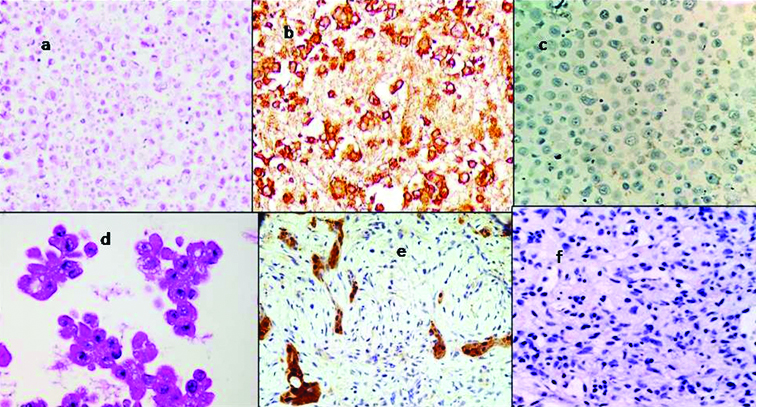
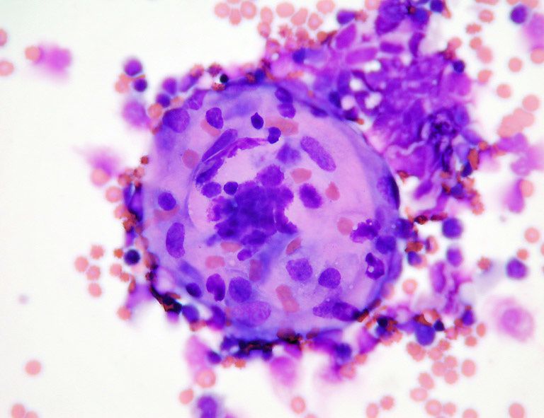
Mesothelial Cells In Peritoneal Fluid, Cytological Diagnosis Of Metastatic Alveolar Rhabdomyosarcoma In The Ascitic Fluid Report Of A Case Highlighting The Diagnostic Difficulties Cytojournal
Mesothelial cells in peritoneal fluid Indeed recently is being sought by users around us, perhaps one of you. People are now accustomed to using the net in gadgets to see video and image information for inspiration, and according to the title of the article I will talk about about Mesothelial Cells In Peritoneal Fluid.
- Mesenchymal Conversion Of Mesothelial Cells Is A Key Event In The Pathophysiology Of The Peritoneum During Peritoneal Dialysis
- Http Www Asl5 Liguria It Portals 0 Anatomiapatologica2015 20150924 Effusion Cytology Pdf
- Fluid Cytology In Serous Cavity Effusions
- Mesothelial Cells Reactive Springerlink
- Reactive Mesothelial Cells Labce Com Laboratory Continuing Education Medical Laboratory Hematology Body Fluid
- Https Encrypted Tbn0 Gstatic Com Images Q Tbn 3aand9gcseb7aizju0qbedevvi8irefmeqt7 Rh3pa 8irkz1p4jn0mu35 Usqp Cau
Find, Read, And Discover Mesothelial Cells In Peritoneal Fluid, Such Us:
- Peritoneal Fluid Veterian Key
- Cytological Diagnosis Of Peritoneal Endometriosis
- Https Encrypted Tbn0 Gstatic Com Images Q Tbn 3aand9gcr S D Awpannkakza 0ldk Epafkthta17xwyo8 N I 9tiksc Usqp Cau
- File Benign Mesothelial Cells Pleural Fluid High Mag Jpg Wikimedia Commons
- Https Www Rcpath Org Asset Ed8cdd8d 8d04 4b82 Ad48d585e2f023be
- Dudley Simmons
- Mickey Mouse Clubhouse Coloring Pages Baby
- Epp Mesothelioma Surgery
- Michael Peters Orthodontist East Lansing
- Pokemon Pictures To Color And Print
If you are looking for Pokemon Pictures To Color And Print you've come to the right place. We have 104 images about pokemon pictures to color and print including images, pictures, photos, wallpapers, and more. In such page, we also provide number of graphics out there. Such as png, jpg, animated gifs, pic art, logo, black and white, translucent, etc.
Furthermore 90k levels correlated to ascitic s il 2r content.

Pokemon pictures to color and print. In addition the fluid within the peritoneal cavity is a battleground in which effector mechanisms generated with the involvement of peritoneal mesothelial cells meet the contaminants. The introduction of peritoneal dialysis pd as a modality of renal replacement therapy has provoked much interest in the biology of the peritoneal mesothelial cell. If the pmn count increases to 250ul or more there are high chances of the presence peritonitis.
In addition to pleura mesothelial cells form a lining around the pericardium the heart and the peritoneum the abdomens inner surface. The amount of fluid is normally small less than 50 ml in humans and contains neutrophils mononuclear cells eosinophils macrophages lymphocytes desquamated mesothelial cells and an average of 30 gml of protein. Such markers of mesothelial cell mass or function that can be measured in the effluent.
Peritoneal mesothelium was found to produce five fold more 90k than ovarian cancer cells. Peritoneal mesothelial cells are in direct contact with the dialysate. Cuboidal mesothelial cells may be found at areas of injury the milky spots of the omentum and the peritoneal side of the diaphragm overlaying the lymphatic lacunae.
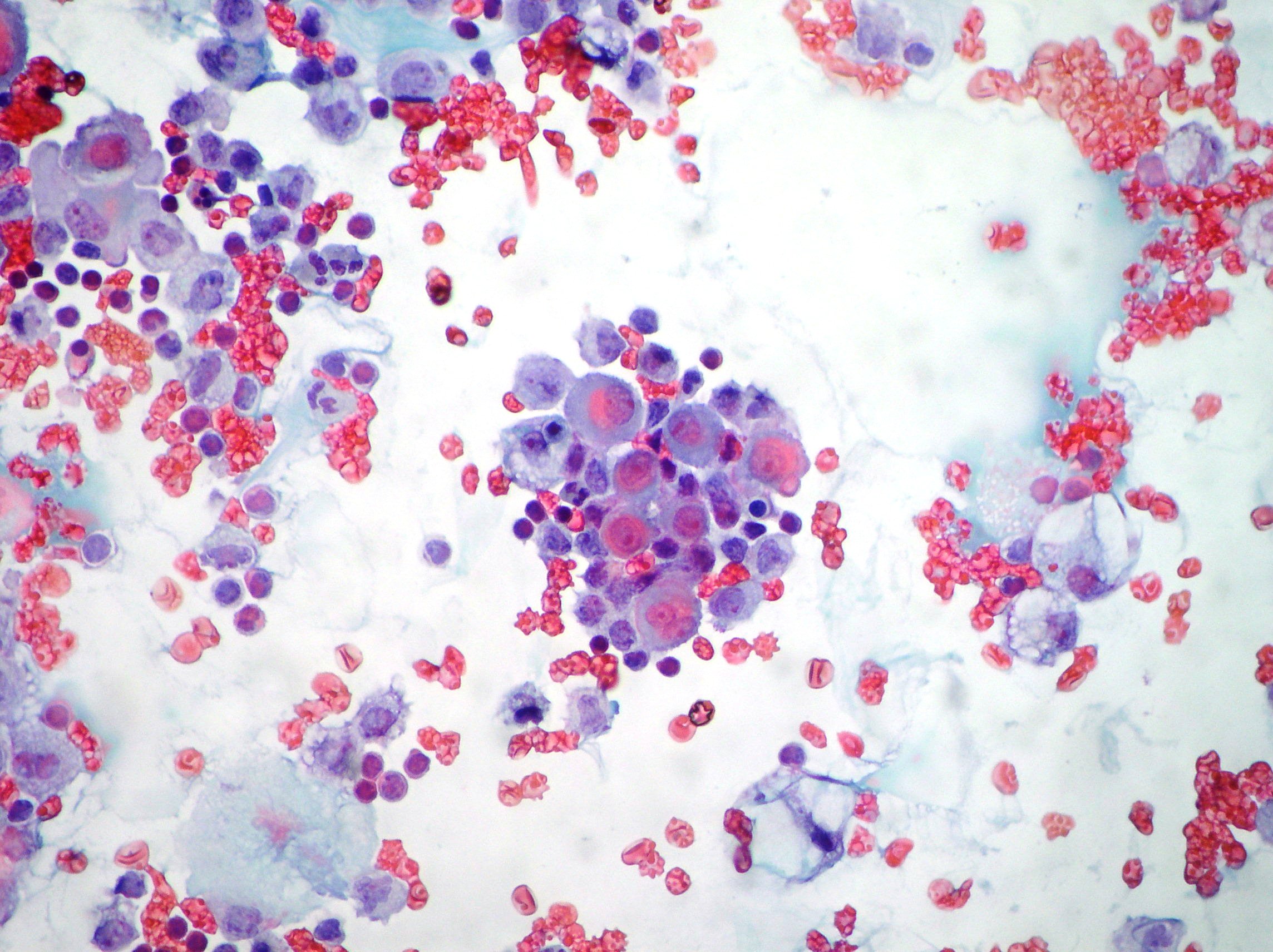
In inflammatory conditions there is a greater number of reactive mesothelial and polys whereas in case of transudate there may be a greater number of lymphocytes. The luminal surface is covered with microvilli. Reactive lymphocytes or plasma cells may be seen.
Mixture of non degenerate neutrophils and macrophages roughly 5050 but can vary from 2080 to 8020 with low numbers of small lymphocytes and mesothelial cells. For the tuberculosis and peritoneal carcinomatosis cases lymphocytes are predominant. The result is a complex mix of cascading processes that have evolved to protect life in the absence of surgery.
The proteins and serosal fluid trapped by the microvilli provide a slippery surface for internal organs to slide past one another. Mesothelial cells isolated from omental tissue have immunohistochemical markers that are identical to those of mesothelial stem cells and omental mesothelial cells can be cultivated in vitro to study changes to their. Mesothelial cells of peritoneal fluid.
Peritoneal fluid is produced by transudation from submesothelial vessels across the peritoneal membrane. Mesothelial cells often appear like squamous cells when microscopically examined however despite their structure that resembles the squamous cells they are a unique form of an epithelial cell. Brownlow ma hutchins dr johnston kg.
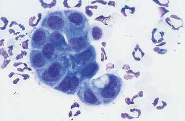
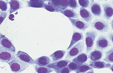
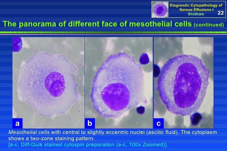
Normal mesothelial cells showed an oval nucleus with finely reticular chromatin and pale blue cytoplasm. Cells in the peritoneal fluid from 159 horses were examined in giemsa stained preparations using light microscopy.
More From Pokemon Pictures To Color And Print
- Wolf Coloring Pages For Girls
- Halloween Party Pictures
- Pumpkin Carving Ideas Easy Scary
- Painted Pumpkin Faces Templates
- Dionne Clueless Costume
Incoming Search Terms:
- Peritoneal Fluid Veterian Key Dionne Clueless Costume,
- Https Www Rcpath Org Asset Ed8cdd8d 8d04 4b82 Ad48d585e2f023be Dionne Clueless Costume,
- Cureus Noninfectious Cloudy Peritoneal Effluent In A Peritoneal Dialysis Patient With Mantle Cell Lymphoma Dionne Clueless Costume,
- Plos One Diagnostic Algorithm For Determining Primary Tumor Sites Using Peritoneal Fluid Dionne Clueless Costume,
- Home Dionne Clueless Costume,
- Https Encrypted Tbn0 Gstatic Com Images Q Tbn 3aand9gcr S D Awpannkakza 0ldk Epafkthta17xwyo8 N I 9tiksc Usqp Cau Dionne Clueless Costume,
