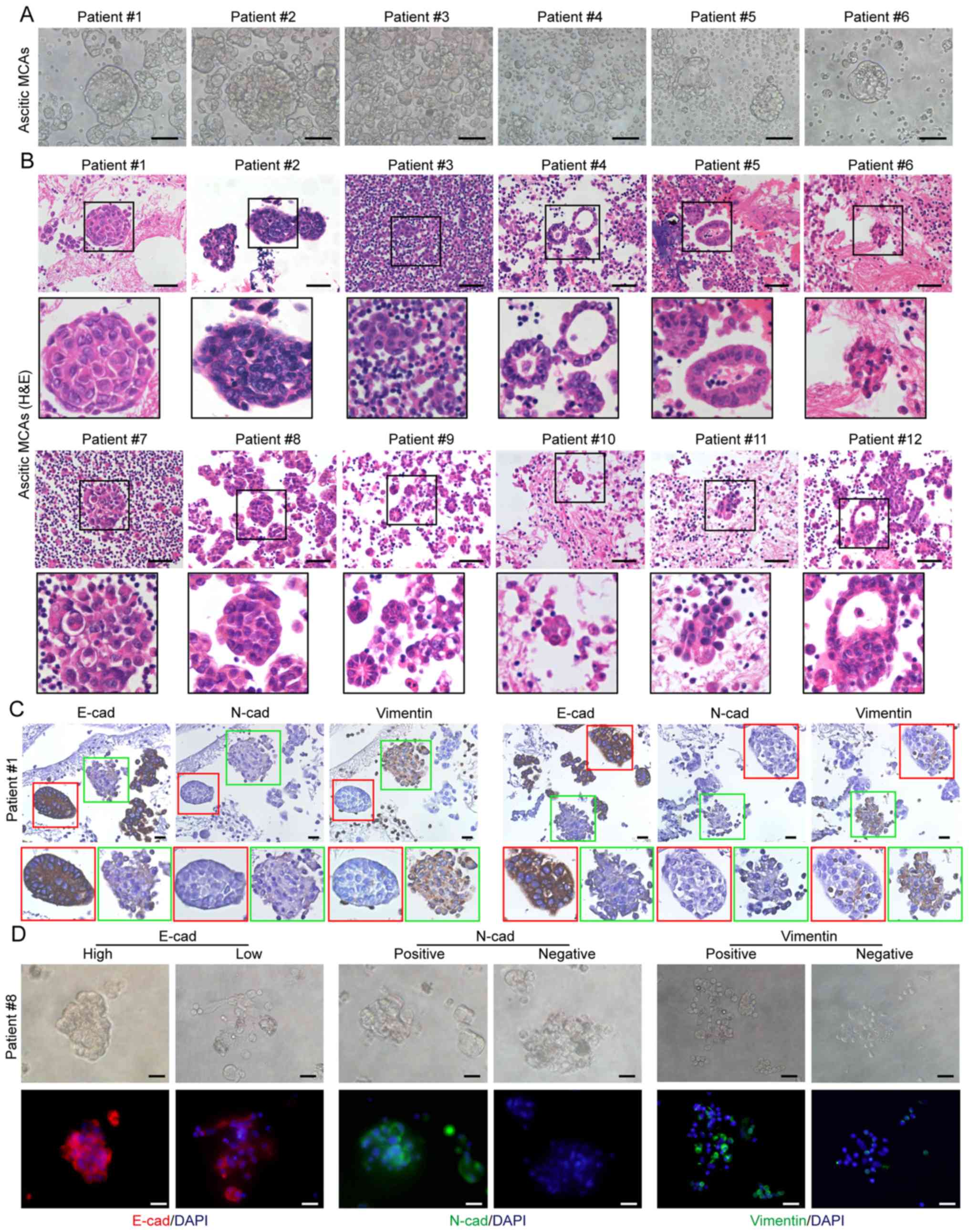
Mesothelial Cells In Ascitic Fluid, The Cell Morphological Characteristics Of Adenocarcinoma Cells Download Scientific Diagram
Mesothelial cells in ascitic fluid Indeed lately has been sought by consumers around us, perhaps one of you. Individuals now are accustomed to using the net in gadgets to view image and video information for inspiration, and according to the name of the post I will talk about about Mesothelial Cells In Ascitic Fluid.
- Oman Medical Journal Archive
- File Benign Mesothelial Cells Pleural Fluid High Mag Jpg Wikimedia Commons
- Cytology Of Pleural And Peritoneal Lesions Chapter 5 Practical Pathology Of Serous Membranes
- Haematology Atlas Of Serous Body Fluids Free Medical Atlas
- Https Encrypted Tbn0 Gstatic Com Images Q Tbn 3aand9gctjxi34atmipfik6awy9fwku9hnngvwavizoylai85qzyd 85bh Usqp Cau
- 01 Presentation I Vs 8 55mb 3 28 08 Pps
Find, Read, And Discover Mesothelial Cells In Ascitic Fluid, Such Us:
- After Extraction Of Ascites Pathology Showing The Ascitic Fluid Download Scientific Diagram
- Cytology Of Body Fluid Pleural Peritoneal Pericardial Ppt Video Online Download
- Https Pathology Ubc Ca Files 2012 06 Fluidcytologybook09r1 Pdf
- Peritoneal Fluid Analysis Litfl Ccc Investigations
- Inhibition Of Hyperglycolysis In Mesothelial Cells Prevents Peritoneal Fibrosis Science Translational Medicine
- What Is Metastatic Mesothelioma
- Diffuse Mesothelioma Payment Scheme Application
- Print And Color Pages
- Easy Darth Vader Coloring Page
- Horse Colorings
If you re searching for Horse Colorings you've arrived at the perfect place. We have 104 graphics about horse colorings including images, photos, pictures, wallpapers, and more. In such web page, we also provide number of images out there. Such as png, jpg, animated gifs, pic art, logo, black and white, translucent, etc.
Cd90 Mesothelial Like Cells In Peritoneal Fluid Promote Peritoneal Metastasis By Forming A Tumor Permissive Microenvironment Horse Colorings
The main purpose of these cells is to produce a lubricating fluid that is released between layers providing a slippery non adhesive and protective surface to facilitate intracoelomic movement.

Horse colorings. Its in vitro release was investigated in primary cultured normal human peritoneal mesothelial cells hpmc. Transudate fluid will be clear and straw color. Abnormal cell morphology ascitic fluid.
C1 and c2 immunohistochemistry showed nuclear positivity for wt1 and nuclear and cytoplasmic staining for calretinin. Ascitic fluid analysis or peritoneal fluid analysis is the major diagnostic test to study the pathophysiology of accumulation of fluid in the peritoneum including diagnosing the causes and inflammation of the fluids. Degenerative mesothelial cells p63 and p40 are very helpful to detect squamous cells cancer cytopathol 2009.
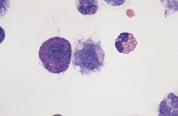
However any fluid that has mesothelial cells with more than 3 nuclei is abnormal and should be sent for hematology or pathology review. To elucidate the source of protein 90k in ascitic fluid. In higher magnification features such as cell in cell engulfment giant atypical mesothelial cells were seen.
Gross or physical appearance. Mesothelial cells in ascitic fluid the associated tumor antigen 90k is known to possess properties similar cytokine in modulating the cellular immune system where accessory cells are the main target of this molecule. Binucleate mesothelial cells seen in image 2 are a normal variant found in any fluid with mesothelial cells.
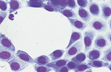
Mesothelial cells the mesothelium forms a monolayer of cells lying on a basement membrane. Ascitic fluid cytomorphologic useless findings cytoplasmic vacuoles signet ring cells individual psammoma bodies. This is seen in blocked lymphatic vessels and the color is milky.
1 it covers both the visceral and parietal peritoneal surfaces. The mesothelium is composed of an extensive monolayer of specialized cells mesothelial cells that line the bodys serous cavities and internal organs. In 67 patients with ovarian cancer with significant amounts of ascites were immune stimulating proteins 90k detected in all.
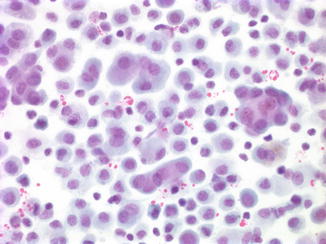
The peritoneal fluid ascites analysis includes. At times its difficult to differentiate between the malignant and mesothelial cellsthe morphology of mesothelial cells. Peritoneal mesothelium was found to produce five fold more 90k than ovarian cancer cells.
No rbcs are seen. White blood cells are 300 cmm. Rarely trinucleate mesothelial cells can be seen.
Furthermore 90k levels correlated to ascitic s il 2r content. Grossly peritoneal fluid is clear and light yellow with 50 ml volume. On cross section the flattened cells are covered.
More From Horse Colorings
- Miguel Coco Costume
- Rapunzel Coloring Pages Printable
- Mesothelioma Prevention After Exposure
- Mesothelioma Asbestos Related Cancer
- Free Easter Coloring Sheets
Incoming Search Terms:
- Mesothelial Cells Review Mesothelial Cells In Ascitic Fluid Free Easter Coloring Sheets,
- Mesothelial Cells In Body Fluid Stock Photo C Toeytoey 99261482 Free Easter Coloring Sheets,
- 8 Mejores Imagenes De Liquidos De Puncion Puncion Hematologia Liquidos Free Easter Coloring Sheets,
- Diagnostic Pitfalls In Effusion Fluid Cytology Basicmedical Key Free Easter Coloring Sheets,
- Https Www Rcpath Org Asset Ed8cdd8d 8d04 4b82 Ad48d585e2f023be Free Easter Coloring Sheets,
- Https Www Rcpath Org Asset Ed8cdd8d 8d04 4b82 Ad48d585e2f023be Free Easter Coloring Sheets,


