Lung Cells Under Microscope, Lung Cells Under The Microscope Ipad Case Skin By Zosimus Redbubble
Lung cells under microscope Indeed recently has been hunted by users around us, perhaps one of you personally. People now are accustomed to using the internet in gadgets to see image and video information for inspiration, and according to the title of the article I will talk about about Lung Cells Under Microscope.
- Human Lung Tissue Under Microscope View Stock Image Image Of Effects Anatomy 101066885
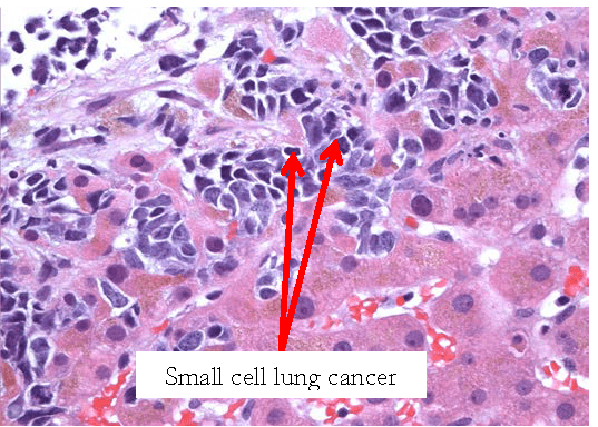
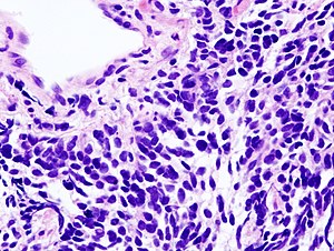
- Small Cell Carcinoma Of The Lung Libre Pathology
- Immune And Inflammatory Cell Composition Of Human Lung Cancer Stroma
- Effect Of Magnetic Fluid Hyperthermia On Lung Cancer Nodules In A Murine Model
- Lung Tissue Bioninja
- Lung
Find, Read, And Discover Lung Cells Under Microscope, Such Us:
- Light Microscopy Of Lung Tissues In Different Groups H E A Showing Download Scientific Diagram
- View Image
- Lung Ageing And Copd Is There A Role For Ageing In Abnormal Tissue Repair European Respiratory Society
- Effect Of Magnetic Fluid Hyperthermia On Lung Cancer Nodules In A Murine Model
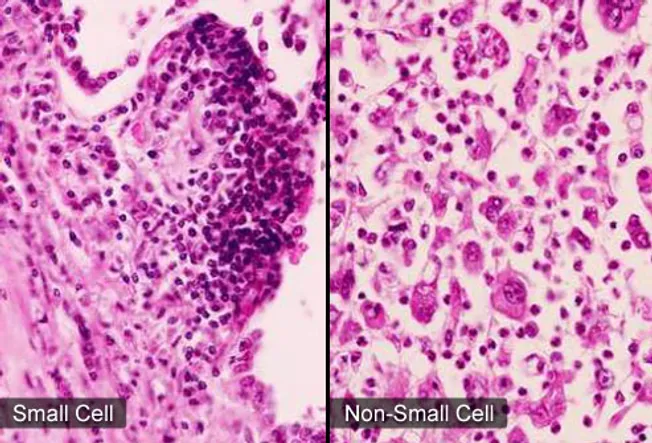
- Non Small Cell Lung Cancer 8 Facts About Nsclc
- Cheap Costumes
- Rudolph The Red Nosed Reindeer Coloring Pages
- Life Expectancy Of Stage 4 Mesothelioma Cancer
- Lung Cancer Staging Mesothelioma
- Masha I Medved Coloring Pages
If you re looking for Masha I Medved Coloring Pages you've reached the right location. We ve got 104 graphics about masha i medved coloring pages including images, photos, pictures, wallpapers, and much more. In these web page, we additionally provide number of graphics out there. Such as png, jpg, animated gifs, pic art, symbol, black and white, translucent, etc.
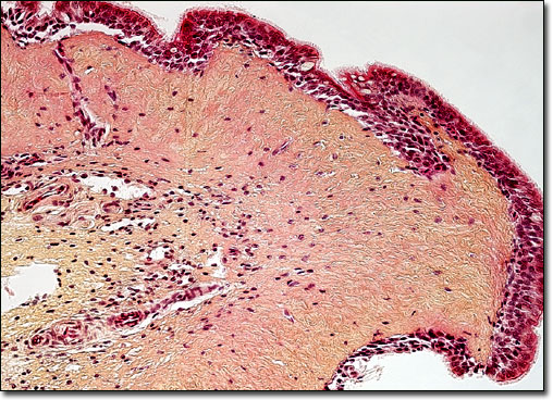
Human lung tissue stained with hematoxylin and eosin as seen under a microscope.
Masha i medved coloring pages. A few epithelial layers are constructed from cells that are said to have a transitional shape. A squamous epithelial cell looks flat under a microscope. Small cell lung cancer.
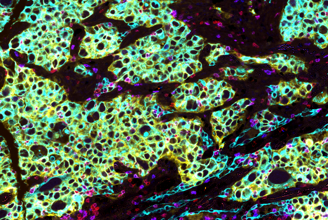
Human lung under the microscope at 100x magnification. Histology help 1 also known as microscopic anatomy or microanatomy 1 is the branch of biology which studies the microscopic anatomy of biological tissues. New detailed images of lung cells under a special microscope illustrate how intense a novel coronavirus infection in the airways can be according to researchers from the university of north.
Targeting lung cancer with a one two punch november 5th 2019 immunotherapies harness a patients own immune system to detect and destroy cancer cells. The two general types of lung cancer include. These lung sections are stained with haematoxylin wiki and eosin wiki which will stain cell nuclei blue and the cell cytoplasm wiki pink.
Your doctor makes treatment decisions based on which major type of lung cancer you have. The lungs are covered by a thin tissue layer called the pleura. A cuboidal epithelial cell looks close to a square.
A thin layer of fluid acts as a lubricant allowing the lungs to slip smoothly as they expand and contract with each breath. The four types of lung cancer are based on the cell types and location as well as how they look under a microscope. When they work they can work wonders.
The healthy human lung seen under a microscope will show typically clear air sacs aveoli wiki. Each lung houses a bronchial tree which gets its name from the intricate network of air passages that supply the lungs with air. This same kind of thin tissue covers the inside of the chest cavity which is also known as pleura.
The air filled sacs in the lungs called alveoli resemble grape clusters. Blood cells known as macrophages located inside each alveolus ingest and destroy airborne irritants that enter the lungs. This cancer often starts in the bronchi major airways.
These will be surrounded by cells of the lung comprising small capillary blood vessels bronchioles and the aveoli. Small cell lung cancer oat cell lung cancer. Doctors divide lung cancer into two major types based on the appearance of lung cancer cells under the microscope.
More From Masha I Medved Coloring Pages
- Poison Ivy Coloring Pages
- Show Me Pictures Of Costumes
- Three Blind Mice Costume
- Snow Tex 16 Oz
- Chameleon Coloring Pages Printable
Incoming Search Terms:
- Alveolar Regeneration Through A Krt8 Transitional Stem Cell State That Persists In Human Lung Fibrosis Nature Communications Chameleon Coloring Pages Printable,
- Understanding Why Lung Cancer Spreads Mit News Massachusetts Institute Of Technology Chameleon Coloring Pages Printable,
- Lung Cancer Pictures X Rays Of Tumors Screening Symptoms And More Chameleon Coloring Pages Printable,
- Https Encrypted Tbn0 Gstatic Com Images Q Tbn 3aand9gcqffb0vasvzfc1zr9o9h Vgh8x Fbbyyagi50dvucpdbpcj3xt Usqp Cau Chameleon Coloring Pages Printable,
- Lung Cells Under The Microscope Stock Photo Image Of Illness Bionic 57194666 Chameleon Coloring Pages Printable,
- Lung Cells Under The Microscope Art Print By Zosimus Redbubble Chameleon Coloring Pages Printable,


