Normal Lung Cells Under Microscope, Crosssection Of Rat Normal Lung Tissue 60x Zoom Stock Photo Download Image Now Istock
Normal lung cells under microscope Indeed recently has been hunted by consumers around us, perhaps one of you. People now are accustomed to using the internet in gadgets to see video and image data for inspiration, and according to the name of the article I will talk about about Normal Lung Cells Under Microscope.
- Crosssection Of Rat Normal Lung Tissue 100x Zoom Stock Photo Download Image Now Istock
- Small Cell Carcinoma Wikipedia
- Normal Lung Microscopic Cells Pathology Lunges
- Autologous Transplantation Of Adipose Derived Mesenchymal Stem Cells Markedly Reduced Acute Ischemia Reperfusion Lung Injury In A Rodent Model Journal Of Translational Medicine Full Text
- Siu Som Histology Crr
- Mic Uk Human Cells Part Ii An Overview For Light Microscopists Lungs
Find, Read, And Discover Normal Lung Cells Under Microscope, Such Us:
- Red Blood Cell Definition Functions Facts Britannica
- Plos Genetics Mir 3607 3p Suppresses Non Small Cell Lung Cancer Nsclc By Targeting Tgfbr1 And Ccne2
- Connective Tissue Lab
- Smoker S Lung Pictures Smokers Lungs Vs Healthy Lungs
- Lung Cells Under The Microscope Stock Photo Image Of Illness Bionic 57194666
- Coloring Activity For Nursery
- Free Halloween Picture Frames
- Mesothelioma Peritoneal Surgery
- Coloring Page Websites
- Sweeney Firm
If you re looking for Sweeney Firm you've arrived at the ideal place. We have 104 graphics about sweeney firm including images, photos, photographs, backgrounds, and much more. In these page, we additionally provide variety of images available. Such as png, jpg, animated gifs, pic art, logo, black and white, transparent, etc.

Concept Of Education Anatomy And Human Lung Tissue Under Microscope Concept Of Spon Anatomy Human Concept Educ Human Lungs Human Tissue Microscopic Sweeney Firm
Non small cell lung cancer homo sapiens 59 yrs caucasian crl 2868 hcc827 lung epithelial adenocarcinoma homo sapiens.

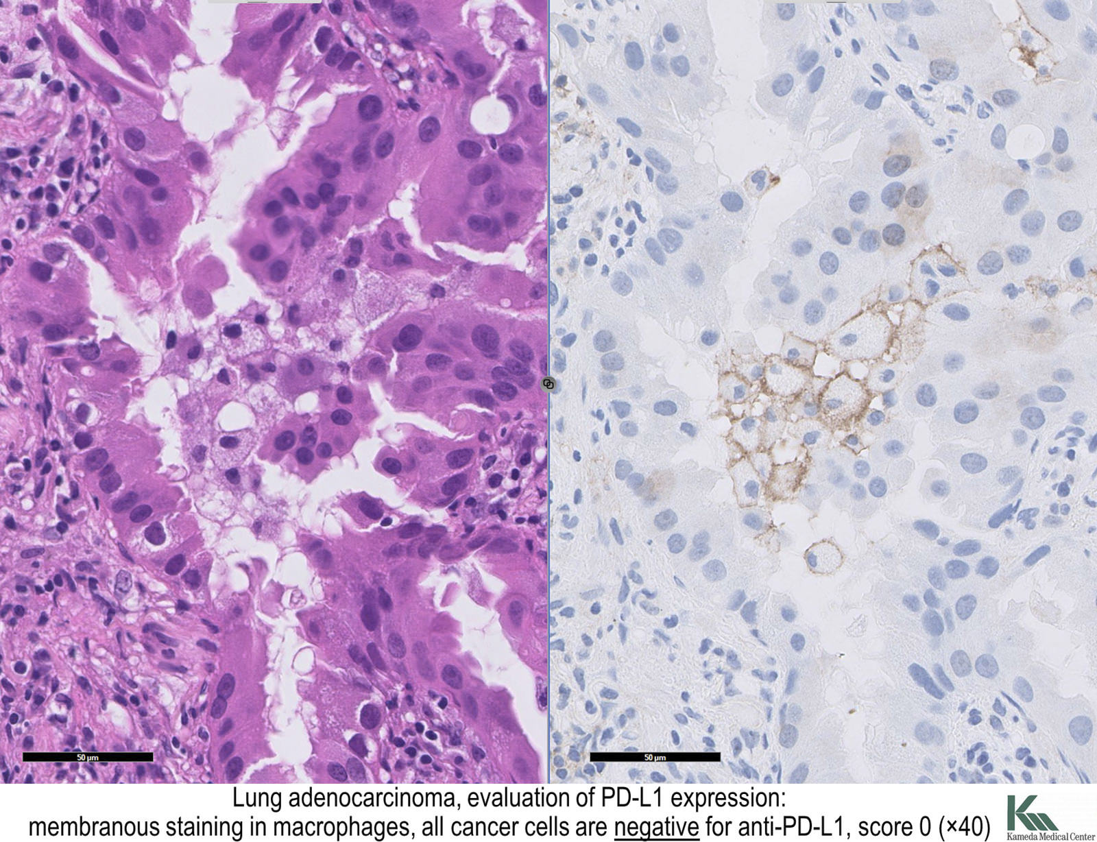
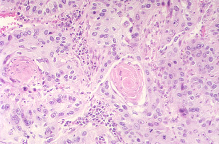
Sweeney firm. Lymph node 3b adenocarcinoma. In contrast to normal cells cancer cells often exhibit much more variability in cell sizesome are larger than normal and some are smaller than normal. Under a microscope normal cells and cancer cells may look quite different.
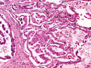
These lung sections are stained with haematoxylin wiki and eosin wiki which will stain cell nuclei blue and the cell cytoplasm wiki pink. The nucleus in cancer cells is rather dark which is. X 100 healthy lung.
What are the alveoli thin tissue sections of lung examined using a microscope give an appearance of a fine lace like structure. Briefly rinse the cell layer with 025 wv trypsin 053 mm edta solution to remove all traces of serum which contains trypsin inhibitor. Cancer cells vary greatly in size they can be of an abnormal shape as well.
Given that they do not attach to each other as other normal cells do in various tissues they also appear as a chaotic collection of cells when viewed under the microscope. A single layer of epithelial cells make up the alveoli. Under a microscope the cells and surrounding tissues become visible as a well appointed city but a city ravaged by the toxic cloud of smoke that has descended upon it.
Lung cancer and normal cell lines atcc no. Cancer cells on the other hand are irregular in shape and misshapen with varying sizes. Add 20 to 30 ml of trypsin edta solution to flask and observe cells under an inverted microscope until cell layer is dispersed usually within 5 to 15 minutes.
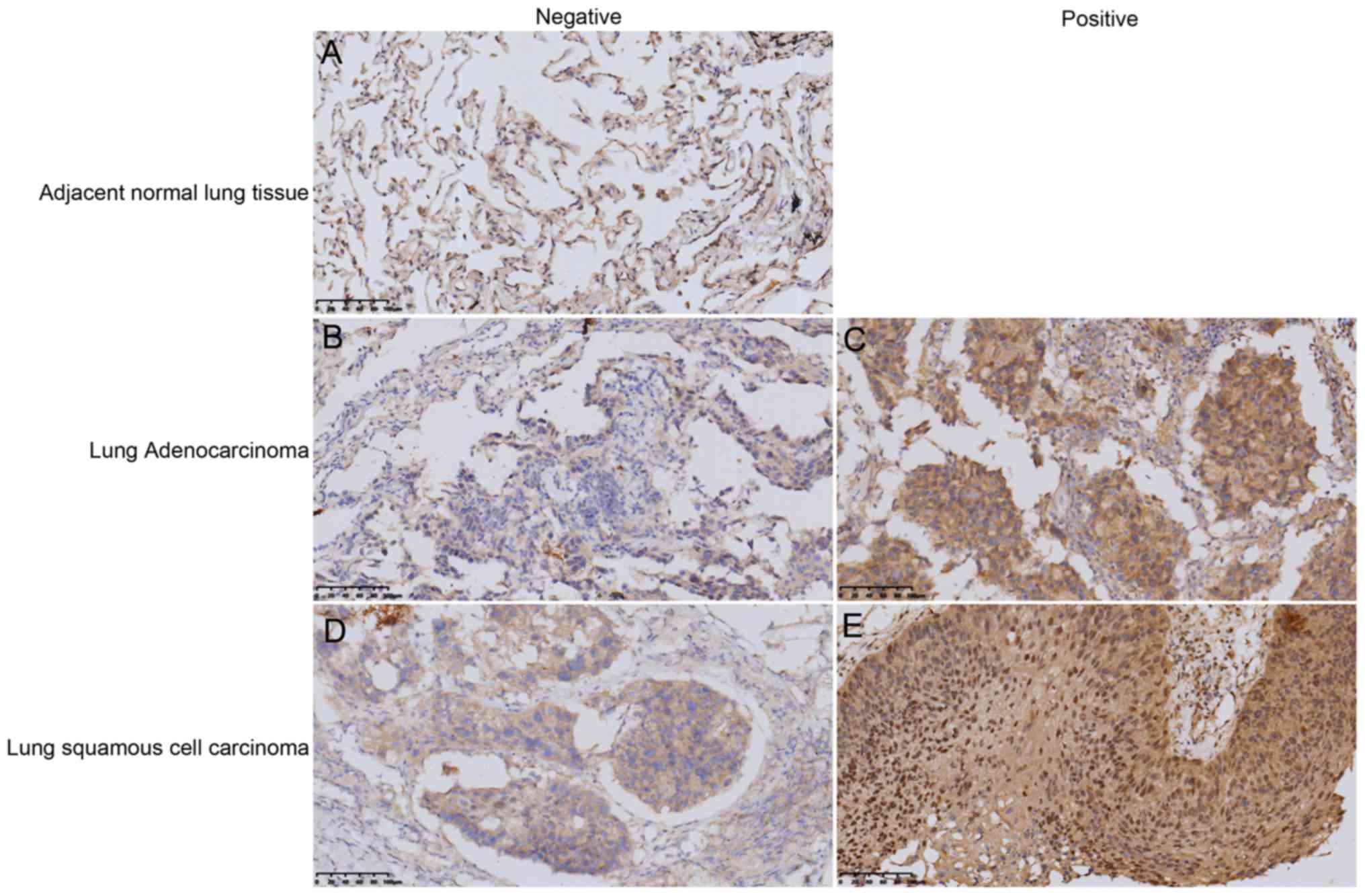
Healthy lung clear air sacs. X400 healthy lung. When viewed under a microscope normal cells are more consistent in their size.
Endothelial cells line alveolar capillaries. For example lung cells remain in the lungs. Histology of normal lung.
The nucleus in the normal cells looks smaller and lighter in normal cells as compared to cancer cells. Human lung under the microscope at 400x magnification. The healthy human lung seen under a microscope will show typically clear air sacs aveoli wikithese will be surrounded by cells of the lung comprising small capillary blood vessels.
Images were captured to an sd card and downloaded to the computer. However most of the normal lung consists of air spaces lined by thin walled alveoli.
More From Sweeney Firm
- Christmas Fairy Coloring Pages
- Decker Law Office
- Riverbank Ice Skating Rink Price
- Real Life Detective Pikachu Coloring Pages
- Christmas Coloring Pages 2018
Incoming Search Terms:
- Human Pathology Nikon S Microscopyu Christmas Coloring Pages 2018,
- What If We Could Stop Lung Cancer Before It Starts School Of Medicine Christmas Coloring Pages 2018,
- Small Cell Carcinoma Wikipedia Christmas Coloring Pages 2018,
- Lung Cells Under The Microscope Stock Photo Image Of Illness Bionic 57194666 Christmas Coloring Pages 2018,
- Mic Uk Human Cells Part Ii An Overview For Light Microscopists Lungs Christmas Coloring Pages 2018,
- Pulmonary Vascular Endothelialitis Thrombosis And Angiogenesis In Covid 19 Nejm Christmas Coloring Pages 2018,


