Electron Microscopy Mesothelioma, Https Cancerres Aacrjournals Org Content 47 12 3199 Full Text Pdf
Electron microscopy mesothelioma Indeed lately is being hunted by users around us, maybe one of you. People are now accustomed to using the internet in gadgets to see image and video data for inspiration, and according to the name of this post I will discuss about Electron Microscopy Mesothelioma.
- Http Vet Sagepub Com Content 20 5 531 Full Pdf
- Malignant Pleural Mesothelioma Pulmonology Advisor
- Pembrolizumab Associated Minimal Change Disease In A Patient With Malignant Pleural Mesothelioma Springerlink
- Detecting Asbestos Using Electron Microscopy
- Malignant Mesothelioma Current Approaches To A Difficult Problem Raja M Flores Md Thoracic Surgery Memorial Sloan Kettering Cancer Center Pdf Free Download
- Wo2017014337a1 Method For Preparing Sample For Scanning Electron Microscopic Observation Of Pathogen Species Including Novel Paraffin Tissue For Diagnosis Of Malignant Mesothelioma Google Patents
Find, Read, And Discover Electron Microscopy Mesothelioma, Such Us:
- Expression Of Matrix Metalloproteinases Tissue Inhibitors Of Metalloproteinase Collagens And Ki67 Antigen In Pleural Malignant Mesothelioma An Immunohistochemical And Electron Microscopic Study Semantic Scholar
- Mesothelioma Microenvironment Image Details Nci Visuals Online
- The Diagnostic Utility Of Immunohistochemistry And Electron Microscopy In Distinguishing Between Peritoneal Mesotheliomas And Serous Carcinomas A Comparative Study Modern Pathology
- Laboratory Diagnosis Of Cancer 1 Histological Methods 2 Cytopathology Fnac Exfoliative 3 Immunohistochemistry Em 4 Molecular Diagnosis 5 Tumor Markers Ppt Download
- 5 04 Pleural Disease Charles Hitchcock Md Flashcards Memorang
- Pittsburgh Malpractice Attorneys
- Paw Patrol Chase Halloween Coloring Pages
- Detailed Thanksgiving Coloring Pages For Adults
- Fashion Coloring Pages Printable
- Childrens Coloring Pages Unicorn
If you re looking for Childrens Coloring Pages Unicorn you've come to the perfect place. We ve got 102 graphics about childrens coloring pages unicorn adding pictures, photos, photographs, wallpapers, and more. In such page, we additionally have variety of graphics out there. Such as png, jpg, animated gifs, pic art, logo, black and white, translucent, etc.
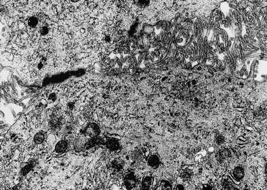
Malignant And Borderline Mesothelial Tumors Of The Pleura Thoracic Key Childrens Coloring Pages Unicorn
This is according to an article published in the journal of thoracic disease titled the use of electron microscopy for diagnosis of malignant pleural mesothelioma electron microscopy which was first developed in the 1930s uses a beam of accelerated electrons to magnify an image of a structure to a much higher degree of magnification than a light microscope.
Childrens coloring pages unicorn. Electron microscopy is a very reliable and useful tool in the diagnosis of certain types of mesothelioma. The localized mesothelioma showed predominance of poorly differentiated cells and fibroblasts. Comin ce de klerk nh henderson dw.
The diagnosis of malignant pleural mesothelioma mpm is challenging and requires immunohistochemistry or electron microscopy assays to specifically differentiate mpm from lung adenocarcinoma. Both light and electron microscopes. An electron microscope is a powerful tool which allows for superior resolution of the specimen with the ability to magnify an object up to two million times.
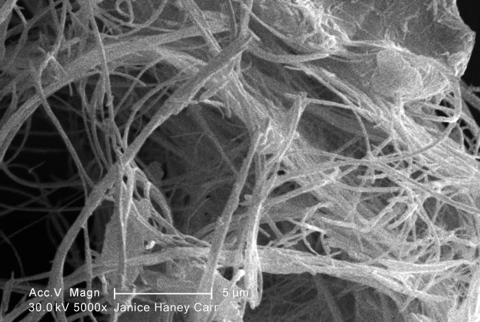
An ultrastructural study of fresh tissue is considered to be the gold standard. Asbestos fibre diameter distributions gauged by electron microscopy were fairly constant irrespective of the degree of fibrosis. Hallmark of epithelioid mesothelioma is the epithelioid cells which are polygonal cells with moderate to abundant eosinophilic cytoplasm vesicular round nuclei and prominent nucleolus.
Current conundrums over risk estimates and whither electron microscopy for diagnosis. The use of electron microscopy for the diagnosis of malignant pleural mesothelioma transmission electron microscopy tem gave a great impulse to medical research. Morphometric examination of desmosomes by electron microscopy may help distinguish epithelial malignant mesotheliomas emm from adenocarcinomas ac.
After its introduction for biological studies in 1931 several microscopic details from observation of animal and tumor cells by tem were published starting from 1953. In patients with cancer involving the lungs liquid often accumulates outside the lungs a feature called pleural effusion. Often mimic nonneoplastic reactive mesothelial cells arch pathol lab med 201814289 the most common histologic patterns of epithelioid mesothelioma are tubulopapillary adenomatoid solid well.
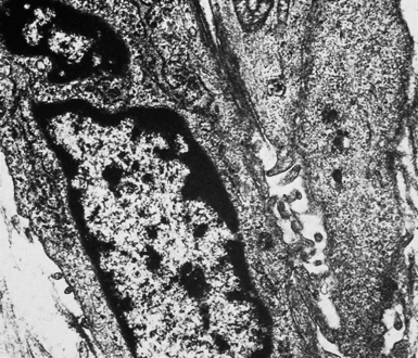
The diffuse mesotheliomas showed features and characteristics of mesothelial cells in the electron microscope whether lining spaces or within solid parts of the tumor. Electron microscopy is already in use to aid the diagnosis of mesothelioma when light microscopy analysis is inconclusive. Electron microscopy is a diagnostic tool using a type of microscopy which utilizes electrons to create an image.
More From Childrens Coloring Pages Unicorn
- Sexy Clown Costume
- Halloween Bulbasaur Coloring Page
- Secret Garden Coloring Pages Pdf
- Hard Gingerbread House Coloring Pages
- Mississippi Mesothelioma Attorney
Incoming Search Terms:
- Extreme Cases Of Asbestos Outbreak News Today Mississippi Mesothelioma Attorney,
- For Diagnosing Mesothelioma Electron Microscopy Still A Valuable Tool Mississippi Mesothelioma Attorney,
- Webpathology Com A Collection Of Surgical Pathology Images Mississippi Mesothelioma Attorney,
- Https Encrypted Tbn0 Gstatic Com Images Q Tbn 3aand9gcrzpsqzi7av H3kot73yhnjt1comrlchw44 Chcwpujk4naswl7 Usqp Cau Mississippi Mesothelioma Attorney,
- Http Informahealthcare Com Doi Pdf 10 3109 01913128809064207 Mississippi Mesothelioma Attorney,
- Microwave Processing References Mississippi Mesothelioma Attorney,
