Mesothelioma Electron Microscopy Tonofilaments, Pathology Outlines Diffuse Malignant Mesothelioma
Mesothelioma electron microscopy tonofilaments Indeed recently is being hunted by users around us, perhaps one of you. People are now accustomed to using the internet in gadgets to see video and image information for inspiration, and according to the name of the post I will talk about about Mesothelioma Electron Microscopy Tonofilaments.
- Https Link Springer Com Content Pdf 10 1007 2f0 387 28274 2 33 Pdf
- Mesothelioma Histology Tonofilaments
- 2
- Tonofibril An Overview Sciencedirect Topics
- Desmosomes Of Epithelial Malignant Mesothelioma
- Https Link Springer Com Content Pdf 10 1007 2f0 387 28274 2 33 Pdf
Find, Read, And Discover Mesothelioma Electron Microscopy Tonofilaments, Such Us:
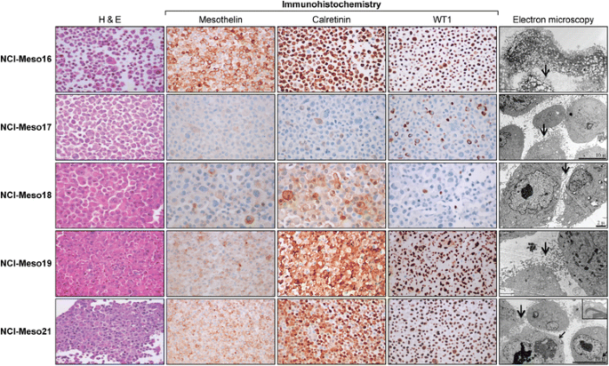
- Mesothelioma Patient Derived Tumor Xenografts With Defined Bap1 Mutations That Mimic The Molecular Characteristics Of Human Malignant Mesothelioma Bmc Cancer Full Text
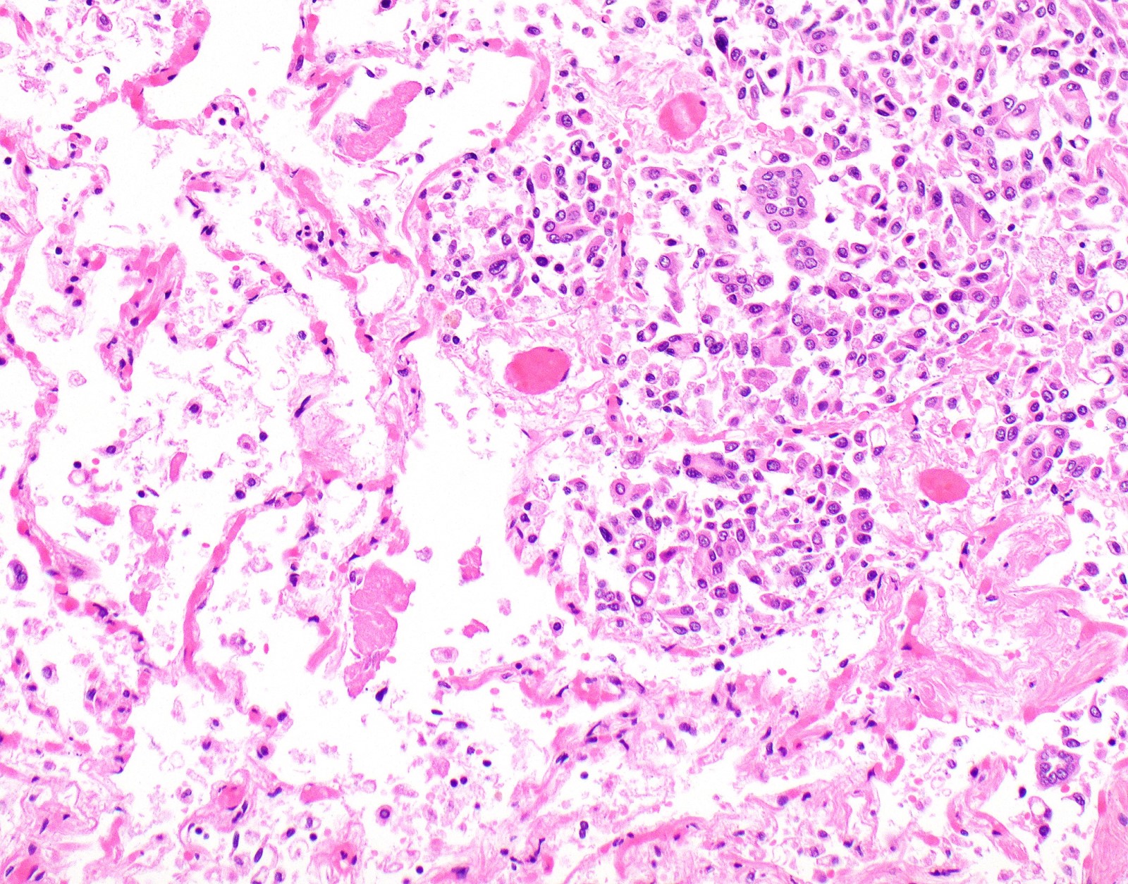
- Pathology Outlines Diffuse Malignant Mesothelioma
- Oa Electron Microscopy Of A Malignant Mesothelial Cell In Tissue From A Download Scientific Diagram
- Pdf Mesothelioma In Domestic Animals Cytological And Anatomopathological Aspects
- Latest Govt Job Bank Recruitment Job Vacancy By Rojgarseva
- Coloring Printables For Preschool
- Talcum Powder Asbestos Mesothelioma
- Mississippi Mesothelioma
- Dot A Dot Art Printables
- Virginia Mesothelioma Doctor
If you are searching for Virginia Mesothelioma Doctor you've come to the right location. We ve got 101 graphics about virginia mesothelioma doctor adding pictures, pictures, photos, wallpapers, and much more. In these web page, we also provide variety of images available. Such as png, jpg, animated gifs, pic art, logo, black and white, translucent, etc.
Https Www Researchgate Net Profile Richard Attanoos Publication 318287816 Guidelines For Pathologic Diagnosis Of Malignant Mesothelioma 2017 Update Of The Consensus Statement From The International Mesothelioma Interest Group Links 5adb02ca458515c60f5cd158 Guidelines For Pathologic Diagnosis Of Malignant Mesothelioma 2017 Update Of The Consensus Statement From The International Mesothelioma Interest Group Pdf Virginia Mesothelioma Doctor
An electron microscopic study of a solitary pleural mesothelioma sarah a.

Virginia mesothelioma doctor. Malignant mesothelioma mm is a neoplasm arising from mesothelial cells lining the pleural peritoneal and pericardial cavities. Alton for secretarial assistance. Lijse md and harlan j.
Cases 1 ant1 2 hat1 similar morphology. T he cell of origin of the solitary pleu ral mesothelioma has been in disputel 411 due in part to the difficulty in proving that the tumors epithelial elements are in. Charbonneau for electron microscopy and to miss m.
But the tissue samples needed for this type of analysis need to be of high quality. Over 20 million people in the us are at risk of developing mm due. Eleven cases of malignant diffuse mesotheliomas histologically classified into two groups epithelial 5 pleural and 3 peritoneal and biphasic or mixed 2 pleural and 1 peritoneal forms were stuied by electron microscopy to elucidate their ultrastructural characteristics.
Malignant mesothelioma and a single small series a. High aspect ratio surface microvilli perinuclear tonofilaments hyaluronic acid crystals. The localized mesothelioma showed predominance of poorly differentiated cells and fibroblasts.
Resembling tonofilaments were present in most tumor cells in different quantity and glycogen. A collection of antibody stains examined under a light microscope can visualize cell structures that differ between the two cancer types. A tonofilament is made up of a variable number of related proteins keratins and is found in all epithelial cells but is particularly well developed in the epidermis.
Radiographic clinical or labo. Mesothelioma of the tunica vaginalis with berep4 and leum1 expression. Mucin positive d pas mucicarmine mesotheliomas aberrant coexpression of adenocarcinoma immunohistochemical markers by mesothelioma or converse c.
Light microscopic analysis was equivocal electron microscopy multiple cytoplasmic tonofilaments and elongated microvilli on the cell surface was used to confirm the diagnosis.
More From Virginia Mesothelioma Doctor
- Multicystic Mesothelioma Peritoneum
- Free Puppy Coloring Pages
- Sexy Girl Coloring Pages
- Lol Doll Free Coloring Pages
- Most Popular Personal Injury Lawyers
Incoming Search Terms:
- Malignant Mesothelioma Electron Microscopy Pdf Free Download Most Popular Personal Injury Lawyers,
- Pathology Outlines Mesothelioma Epithelioid Most Popular Personal Injury Lawyers,
- Https Www Sciencedirect Com Science Article Pii S0031302516351078 Pdf Md5 E4dd5015175a552eff7a24d31ae34874 Pid 1 S2 0 S0031302516351078 Main Pdf Most Popular Personal Injury Lawyers,
- Pathology Outlines Diffuse Malignant Mesothelioma Most Popular Personal Injury Lawyers,
- Pdf Deciduoid Peritoneal Mesothelioma In A Dog Most Popular Personal Injury Lawyers,
- Pathology Outlines Diffuse Malignant Mesothelioma Most Popular Personal Injury Lawyers,


