Diaphragmatic Pleural Calcification, Diseases Of The Chest Wall Pleura And Diaphragm Springerlink
Diaphragmatic pleural calcification Indeed lately has been sought by users around us, perhaps one of you. People are now accustomed to using the net in gadgets to view image and video information for inspiration, and according to the title of the post I will talk about about Diaphragmatic Pleural Calcification.
- Asbestos Plaques Radiology At St Vincent S University Hospital
- Benign Pleural Thickening Radiology Key
- Next
- Pleural Plaques Causes Symptoms Diagnosis Treatment
- Chest X Ray Showing Moderate Right Sided Pleural Effusion And Bilateral Download Scientific Diagram
- Calcified Pleural Plaques Radiology Case Radiopaedia Org
Find, Read, And Discover Diaphragmatic Pleural Calcification, Such Us:
- Calcifications Are Observed Bilaterally At The Level Of Diaphragmatic Download Scientific Diagram
- Pulmonary Tuberculosis Complicating Asbestosis Hkmj
- View Image
- Https Www Ajronline Org Doi Pdf 10 2214 Ajr 115 3 473
- Https Encrypted Tbn0 Gstatic Com Images Q Tbn 3aand9gctyiozt02xlfpon8aohtpg5rrs1qnouulldjojo9fg Usqp Cau
- Peppa Pig Coloring Book Printable
- Coco Coloring Pages Pepita
- Lawyer For Asbestos Exposure
- Mesothelioma Testis Radiology
- Green Bay Personal Injury Mesothelioma Asbestos Lawyers
If you re searching for Green Bay Personal Injury Mesothelioma Asbestos Lawyers you've come to the ideal location. We ve got 104 images about green bay personal injury mesothelioma asbestos lawyers adding images, pictures, photos, backgrounds, and more. In such web page, we also provide number of images out there. Such as png, jpg, animated gifs, pic art, symbol, black and white, translucent, etc.

Focal Pleural Tumorlike Conditions Nodules And Masses Beyond Mesotheliomas And Metastasis Sciencedirect Green Bay Personal Injury Mesothelioma Asbestos Lawyers

Pleura Chest Wall And Diaphragm Chest Radiology The Essentials 2nd Edition Green Bay Personal Injury Mesothelioma Asbestos Lawyers

Pdf Tumorlike Conditions Of The Pleura Green Bay Personal Injury Mesothelioma Asbestos Lawyers
Https Encrypted Tbn0 Gstatic Com Images Q Tbn 3aand9gctm5kutme0w8fvpuuz9gbikhyktiocs1 E5abvzt3c 39dm6eek Usqp Cau Green Bay Personal Injury Mesothelioma Asbestos Lawyers

Pictorial Essay Of Radiological Features Of Benign Intrathoracic Masses Green Bay Personal Injury Mesothelioma Asbestos Lawyers
2 Green Bay Personal Injury Mesothelioma Asbestos Lawyers
This shape is not uncommon.

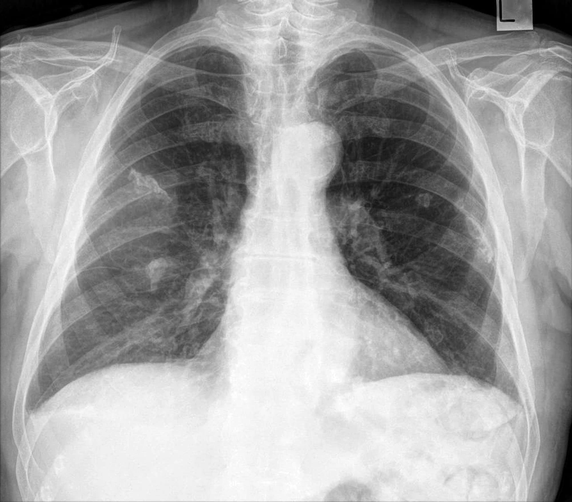
Green bay personal injury mesothelioma asbestos lawyers. What is the difference between calcified and noncalcified pleural plaque. Typically with sparing of the costophrenic angles. Note the thin layer of extrapleural fat arrows that separates the plaques from the underlying rib and intercostal muscle.
Infection involving the pleura. The appearance of plaques has been likened to that of a holly leaf with thickened rolled and nodular edges 4. The plaques were found by histologic six patients and ultrastructural examination two patients to consist of pure collagen.
Non calcified pleural plaques and pleural implants are often better demonstrated with ct rather than by radiography in contrast to pleural effusion or pneumothorax. Locations most commonly encountered include posterolateral mediastinal and diaphragmatic pleura 1. C axial ct image shows nodular calcified plaques.
Calcified plaques are more obvious than non calcified plaques to be identified. All are intimately associated with each other which occasionally makes it difficult to determine the origin of a mass or disease process involving one or more of these compartments disorders involving the chest wall pleura or diaphragmresulting in a pleural based masscan arise from one of these compartments an. Studies have also shown a higher incidence of smokers among those with pleural plaques although research is inconclusive whether smoking played a part in developing the plaques.
Clinical and morphologic observations are described of diaphragmatic and pleural plaques in six patients. Although the exact cause of the plaques is unknown the frequent finding by other investigators of asbestos bodies in the lungs of patients with these plaques. Pleural disease is manifest by the accumulation of fluid or air in the pleural space by pleural thickening with or without calcification or by the presence of a pleural mass.
B coronal ct image shows calcified discrete pleural plaques arrows located along the diaphragm and the lateral pleural surface. Pyothoraxempyema tuberculous pleuritis 3. Calcified pleural plaques from asbestos exposure.
Pleural effusion a number of different types of fluid may accumulate in the pleural space the most common being transudate exudate thin or thick blood and chyle. The pleural calcification that arises from asbestos exposure commonly occurs along the diaphragm and can be bilateral. A small hemithorax with extensive pleural calcification fibrothorax may be seen after tuberculosis and untreated hemothorax.
Pleural calcification can be the result of a wide range of pathology and can be mimicked by a number of conditionsartifacts. The chest wall pleura and diaphragm enclose the outer lung. The plaques commonly develop near the ribs vertebrae or diaphragm.

Radiological Review Of Pleural Tumors Abstract Europe Pmc Green Bay Personal Injury Mesothelioma Asbestos Lawyers

Pleural Plaques Diffuse Pleural Thickening Rounded Atelectasis And Download Scientific Diagram Green Bay Personal Injury Mesothelioma Asbestos Lawyers
Pleural Plaques Test Findings Medschool Green Bay Personal Injury Mesothelioma Asbestos Lawyers

Pleural Calcifications Green Bay Personal Injury Mesothelioma Asbestos Lawyers
More From Green Bay Personal Injury Mesothelioma Asbestos Lawyers
- Princess Tiana Coloring Pages
- Simple Scary Pumpkin Designs
- Sonic Super Smash Bros Coloring Pages
- Rodrigo Sanchez Attorney New York
- Cdc Mesothelioma
Incoming Search Terms:
- Benign Pleural Thickening Radiology Key Cdc Mesothelioma,
- View Image Cdc Mesothelioma,
- Asbestos Related Diseases Wikipedia Cdc Mesothelioma,
- Http Www Lungcancerjournal Info Article S1556 0864 15 31680 4 Pdf Cdc Mesothelioma,
- Diseases Of The Chest Wall Pleura And Diaphragm Springerlink Cdc Mesothelioma,
- Calcified Pleural Plaque Radiology Case Radiopaedia Org Cdc Mesothelioma,
