Xray Benign Lung Tumor, Evaluation Of The Solitary Pulmonary Nodule American Family Physician
Xray benign lung tumor Indeed lately is being sought by consumers around us, perhaps one of you personally. People now are accustomed to using the internet in gadgets to view image and video data for inspiration, and according to the name of this post I will talk about about Xray Benign Lung Tumor.
- Lung Cancer Oncology Medbullets Step 2 3
- What Are Pulmonary Nodules Inogen
- Learningradiology Hamartoma Of Lung
- Pet Ct Imaging In Lung Cancer Indications And Findings
- Solitary Pulmonary Nodule In An 84 Year Old Man Photo Quiz American Family Physician
- The Radiology Assistant Benign Versus Malignant
Find, Read, And Discover Xray Benign Lung Tumor, Such Us:
- Management Of Incidental Lung Nodules 8 Mm In Diameter Sanchez Journal Of Thoracic Disease
- Por Chest X Ray Showing The Left Upper Lobe Lung Mass Download Scientific Diagram
- Lung Cancer Chest X Ray Wikidoc
- Asymptomatic Pulmonary Nodules In A Patient With Early Stage Breast Cancer Cryptococcus Infection Sciencedirect
- Slow Growing Lung Cancer As An Emerging Entity From Screening To Clinical Management European Respiratory Society
- Sonic Coloring Page
- Preschool Daniel And The Lions Den Coloring Page
- Family Pumpkin Painting Ideas
- Kawaii Cute Coloring Pages
- Descendants Coloring Pages Mal And Evie
If you re looking for Descendants Coloring Pages Mal And Evie you've arrived at the right location. We ve got 104 images about descendants coloring pages mal and evie adding images, pictures, photos, backgrounds, and much more. In these web page, we also have number of images out there. Such as png, jpg, animated gifs, pic art, symbol, blackandwhite, translucent, etc.
Swensen et al radiology 2005235259 265.

Descendants coloring pages mal and evie. Types of benign lung tumors include hamartomas adenomas and papillomas. Patients presenting with symptoms and found to have tumors on chest x ray films or computed tomography ct imaging studies are more likely to have a lung malignancy. In fact a nodule shows up on about one in every 500 chest.
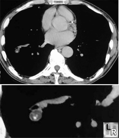
Tumors of the lung 91 hamartoma hamartomas synonyms. Chondroid hamartoma chondromatous hamartoma chondrohamartoma hamartochondroma chondroma are the most common type of benign lung tumor and account for 8 of all lung neoplasms. In almost all cases benign lung tumors require no treatment but your doctor will probably monitor your tumor for changes.
6 tumors and tumor like lesions of the lung the solitary pulmonary nodule spn a solitary pulmonary nodule is defined as a round or oval opacity less than 3 cm in diameter which is surrounded completely by pulmonary parenchyma and is not associated with lymph node enlargement atelectasis or pneumonia midthun et al. A chest x ray alone cannot confirm if a lung nodule mass shadow neoplasm or lesion is cancer or something more benign like a cyst or scar. Benign tumors may also present primarily in the airway versus the lung parenchyma and certain causes may present in either location or in both locations.
A lung tumor is an abnormal rate of cell division or cell death in lung tissue or in the airways that lead to the lungs. Obscured images overlapping structures can obscure tumors on an x ray and make them hard to visualize especially if they are small. Using first follow up diagnostic ct to differentiate benign and malignant lesions.
Indeterminate solitary pulmonary nodules revealed at population based ct screening of the lung. What are benign lung nodules and benign lung tumors. Shodayu takashima et al.
They can be divided into benign and malignant tumors and into those which arise in the ribcage and those of soft tissue density. Benign lung tumors can be classified pathologically but a clinically useful classification would combine location ie endobronchial or parenchymal and information about whether the lesions are. Tumors of the chest wall are varied some of which are found most often in this region.
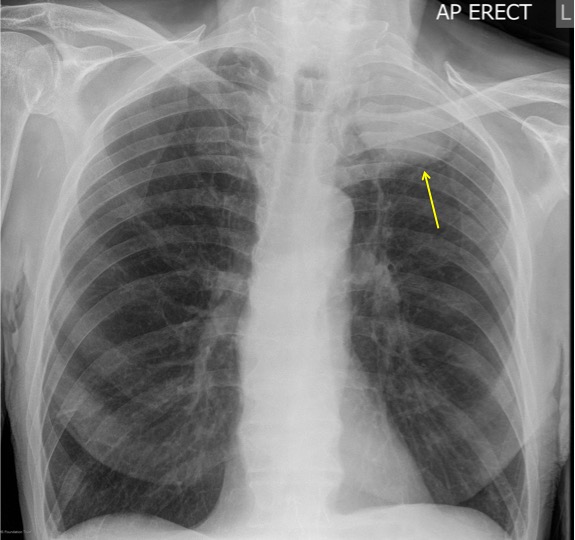
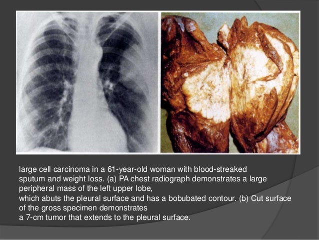
Lung tumors can be seen on a chest x ray when they reach about 1 cm in diameter. A chest x ray is frequently the first test ordered and may pick up a suspicious finding. A nodule is a spot on the lung seen on an x ray or computed tomography ct scan.
Its important to note that a chest x ray alone cannot prove conclusively that a tumor is benign or malignant. As a malformation tumor the hamartoma may contain fatty tissue cartilage epithelial tissue and connective tissue.
More From Descendants Coloring Pages Mal And Evie
- Cat Valentine Coloring Pages
- Easy Pumpkin Carving Ideas For Beginners
- Parker Meyers
- Pumpkin Pictures To Color Free
- Simpson Lawrence Windlass Manual
Incoming Search Terms:
- Carcinoid Lung Tumour Something About Radiology Just For Sharing Simpson Lawrence Windlass Manual,
- Possible Causes Of A Lung Mass Simpson Lawrence Windlass Manual,
- What To Do With Lung Nodules Pulse Today Simpson Lawrence Windlass Manual,
- Abnormal Shadow On Chest Radiograph Medpage Today Simpson Lawrence Windlass Manual,
- Pancoast Syndrome Risk Factors Symptoms And More Simpson Lawrence Windlass Manual,
- How Lung Cancer Is Diagnosed Simpson Lawrence Windlass Manual,
:max_bytes(150000):strip_icc()/lung-mass-possible-causes-and-what-to-expect-2249388-5bc3f847c9e77c00512dc818.png)
/multiple-lung-nodules-causes-and-diagnosis-2249390-v1-5c1ac161c9e77c000122ea44.png)


