Wt1 Immunohistochemistry Mesothelioma, Mesothelioma Positive For Pan Ck Ck 5 6 Calretinin Wt1 Diagnostic Biosystems Immunohistochemistry Primary Antibodies Monoclonal Antibodies Polyclonal Antibodies Fitc Antibodies Automated Staining Instrumentation Mouse And Rabbit
Wt1 immunohistochemistry mesothelioma Indeed recently is being sought by users around us, maybe one of you. People now are accustomed to using the net in gadgets to see video and image information for inspiration, and according to the name of this post I will discuss about Wt1 Immunohistochemistry Mesothelioma.
- 2
- Applied Sciences Free Full Text Immunohistochemical Expression Of Wilms Tumor 1 Protein In Human Tissues From Ontogenesis To Neoplastic Tissues Html
- 2
- Assaybiotechnology
- Ae00126 Aeonian Biotech
- Malignant Mesothelioma And Other Primary Pleural Tumors Thoracic Key
Find, Read, And Discover Wt1 Immunohistochemistry Mesothelioma, Such Us:
- Anti Wt1 Wilms Tumor 1 Antibody Mouse Anti Human Monoclonal Lsbio
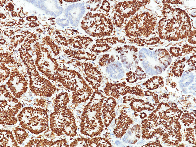
- Mesothelioma Positive For Pan Ck Ck 5 6 Calretinin Wt1 Diagnostic Biosystems Immunohistochemistry Primary Antibodies Monoclonal Antibodies Polyclonal Antibodies Fitc Antibodies Automated Staining Instrumentation Mouse And Rabbit
- Frozen Sections Of Tumors Stained With Anti Wt1 Mab Frozen Download Scientific Diagram
- Anti Wt1 Antibody Mouse Anti Human Wilms Tumor 1 Wt1 Monoclonal Antibody Clone Abt Wt1 Np 077742 2
- Esbe Scientific Wt1 6f H2
- Printable Coloring Books For Kids
- Dr John D Osteraas Mesothelioma
- Poinsettia Flower Coloring Page
- Color By Number Printables For Adults
- Mesothelioma Immunohistochemistry Diagnosis
If you re searching for Mesothelioma Immunohistochemistry Diagnosis you've arrived at the perfect place. We ve got 100 graphics about mesothelioma immunohistochemistry diagnosis including images, pictures, photos, wallpapers, and more. In such web page, we additionally provide variety of graphics available. Such as png, jpg, animated gifs, pic art, symbol, black and white, translucent, etc.
89 mesothelin 132 of 150.
Mesothelioma immunohistochemistry diagnosis. 88 and d2 40 78 of 97. Therefore it is evident that wt1 is the most useful positive marker for the pathological diagnosis of epithelioid mesothelioma. 100 wt1 205 of 218.
Wt1 mutation in malignant mesothelioma and wt1 immunoreactivity in relation to p53 and growth factor receptor expression cell type transition and prognosis j pathol 1811. The best discriminators among the antibodies considered to be negative markers for mesothelioma are cea moc 31 ber ep4 bg 8 and b723. However when compared with calretinin wt1 reactivity tends to be within a limited area graded as 1 or 2.
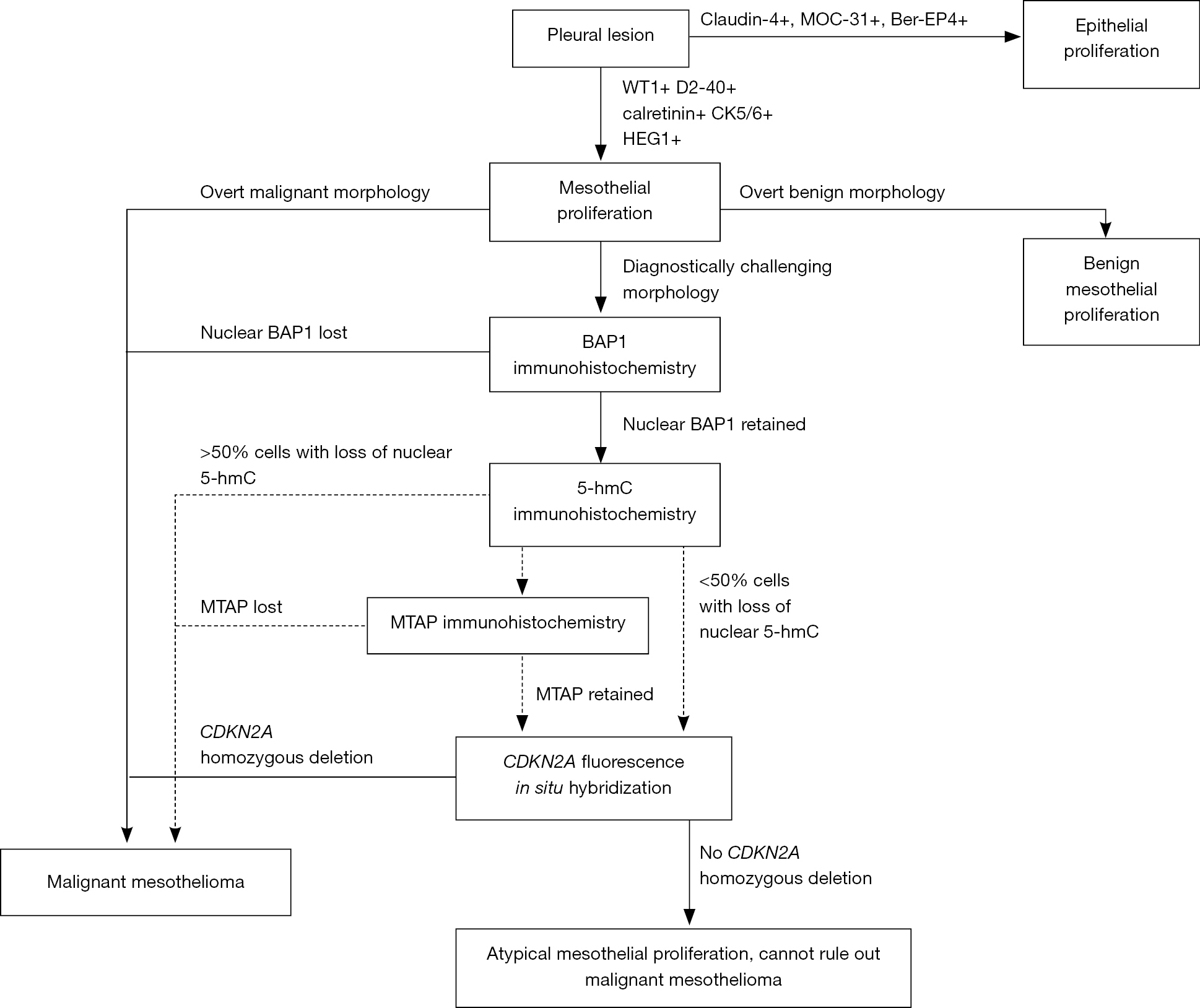
To distinguish malignant mesothelioma adenocarcinoma and reactive benign mesothelium with cytological and histological methods including immunocytochemistry is a major diagnostic challenge. The results of immunohistochemistry markers performed were tabulated. Electron microscopy had been performed on 16 cases.
Wt1 is one of the most useful markers for identifying mesothelioma. Calretinin is a calcium binding protein that occurs in various types of cells in the body. An assessment of recently described markers for mesothelioma and adenocarcinoma.
In the present study the expression of wt1 was examined in 494 cases of human cancers including tumors of the gastrointestinal and pancreatobiliary system urinary tract male and female genital organs breast lung brain skin soft tissues and bone by immunohistochemistry using polyclonal c 19 and monoclonal 6f h2 antibodies against. In addition wt1 had the highest sensitivity and specificity among all positive markers. Nuclear staining for wt1 is highly specific for.
The wilms tumour susceptibility gene 1 wt1 expressed during transition of mesenchyme to epithelial tissues is regarded as a marker for the. This diagnostic technique is called immunohistochemistry. Acute myeloid leukemia cystic partially differentiated nephroblastoma desmoplastic small round cell tumor malignant mesothelioma metanephric adenoma nephrogenic rests ovarian carcinomas serous carcinoma almost all transitional small cell am j surg pathol 2005291034 peritoneal serous carcinoma involving an endometrial polyp 80 am j surg pathol.
Stains molecular markers wt1. 94 ck56 173 of 194. Wt1 may also play a role in making mesothelioma cells resistant to chemotherapy according to a 2017 study in pathology oncology research.
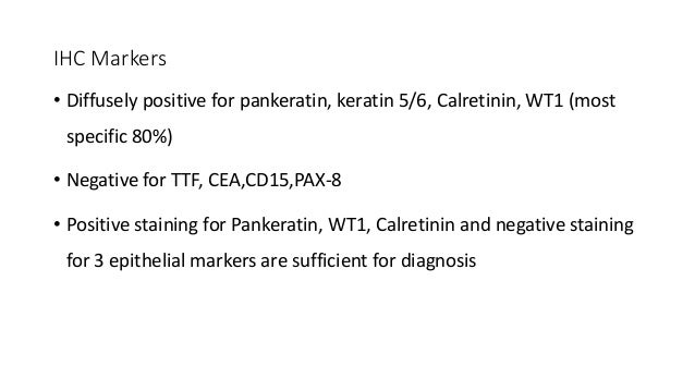
Hbme 1 moc 31 wt1 and calretinin. After analyzing the results it is concluded that calretinin cytokeratin 56 and wt1 are the best positive markers for differentiating epithelioid malignant mesothelioma from pulmonary adenocarcinoma. Immunohistochemistry for mesothelioma is still developing as a science and different pathologists have experience with using different antibodies.
The diagnosis of adenocarcinoma was based on typical light microscopic findings and a positive stain for mucin.
More From Mesothelioma Immunohistochemistry Diagnosis
- Mesothelioma Of Pleural Membrane
- Family Services Lawyer
- Mickey Halloween Images
- Printable Coloring Worksheets For Grade 1
- Myers Pediatric Dentistry
Incoming Search Terms:
- Wilms Tumor 1 Cytokeratin Dual Color Immunostaining Reveals Distinctive Staining Patterns In Metastatic Melanoma Metastatic Carcinoma And Mesothelial Cells In Pleural Fluids An Effective First Line Test For The Workup Of Malignant Effusions Conner Myers Pediatric Dentistry,
- Wt1 Antibody 258 From Biocare Medical Biocompare Com Myers Pediatric Dentistry,
- Anti Wt1 Antibody Rabbit Wilms Tumor 1 Wt1 Monoclonal Antibody Clone Wt1 1434r Np 000369 3 Myers Pediatric Dentistry,
- Mesothelioma Positive For Pan Ck Ck 5 6 Calretinin Wt1 Diagnostic Biosystems Immunohistochemistry Primary Antibodies Monoclonal Antibodies Polyclonal Antibodies Fitc Antibodies Automated Staining Instrumentation Mouse And Rabbit Myers Pediatric Dentistry,
- Anti Wt1 Antibody Mouse Wilms Tumor 1 Wt1 Monoclonal Antibody Clone Rwt1 857 Np 000369 3 Myers Pediatric Dentistry,
- The Diagnostic Utility Of Pax8 Immunostaining Of Malignant Peritoneal Mesothelioma Presenting As Serous Ovarian Carcinoma A Single Center Report Of Two Cases Myers Pediatric Dentistry,


.jpg)

