Trapped Lung Radiology, Pictorial Review Of Non Traumatic Thoracic Emergencies In The Pediatric Population Egyptian Journal Of Radiology And Nuclear Medicine Full Text
Trapped lung radiology Indeed lately has been sought by users around us, perhaps one of you personally. People are now accustomed to using the internet in gadgets to see video and image data for inspiration, and according to the name of the article I will discuss about Trapped Lung Radiology.
- Non Expandable Lung An Underappreciated Cause Of Post Thoracentesis Basilar Pneumothorax Bmj Case Reports
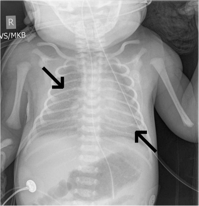
- Pictorial Review Of Non Traumatic Thoracic Emergencies In The Pediatric Population Egyptian Journal Of Radiology And Nuclear Medicine Full Text
- A Systematic Approach To Chest Radiographic Analysis Springerlink
- Slide 1
- Consolidated Lung Emory School Of Medicine
- Pleural Calcifications
Find, Read, And Discover Trapped Lung Radiology, Such Us:
- Pneumothorax Ex Vacuo Following Thoracentesis For Persistent Pleural Effusion Scitechnol
- Pneumothorax Ex Vacuo Or Trapped Lung In The Setting Of Hepatic Hydrothorax Bmc Pulmonary Medicine Full Text
- 60 Imaging Ideas Image Radiology Pleural Effusion
- View Of Loculated Pneumothorax With A Deep Sulcus Sign The Southwest Respiratory And Critical Care Chronicles
- Trapped Lung Eurorad
- Free Fun Coloring Pages
- Malignant Mesothelioma Pathology Staging
- Free Bible Coloring Pages Printable
- Halloween Bride
- Puppy Coloring Pages For Adults
If you re looking for Puppy Coloring Pages For Adults you've reached the ideal location. We have 104 graphics about puppy coloring pages for adults including images, photos, pictures, backgrounds, and more. In these webpage, we also provide variety of images available. Such as png, jpg, animated gifs, pic art, logo, blackandwhite, translucent, etc.
Today is world radiography day and the international day of radiology free video.

Puppy coloring pages for adults. Jeffrey albores md and tisha wang md. A 46 year old woman with end stage liver disease complicated by recurrent hepatic hydrothorax requiring multiple. Notice how the prior pre evacuation pleural effusion causing passive atelectasis mimics the shape of post evacuation.
The elevated total pleural fluid protein may be related to factors other than active pleural inflammation or malignancy and does not exclude the diagnosis. Trapped lung occurs as a mature fibrous strip encircles the visceral pleura restricting the lung expansion which develops from inflammatory sequelae. The term is similar but not entirely synonymous with trapped lung which is due to pleural inflammation from remote disease resulting in fibrous thickening of the pleura.
They underwent a thoracentesis 5 days earlier and in this context these findings represent trapped lung. Trapped lung list of authors. Trapped lung develops as a sequela of pleural space inflammation from remote disease resulting in the development of a mature fibrous membrane that impedes the lung from re expanding.
Conclusionstrapped lung is a clinical entity characterized by the presence of a restrictive visceral pleural peel that was first described in 1967the pleural fluid is paucicellular ldh is low and protein may be in the exudative range. This patient has features of trapped lung on chest x ray and ct not shown which occurs post pleural tap. Trapped lung does not appear larger on expiration than on inspiration in comparison to pneumothorax.
The visceral pleural line delineates the scarred lung contour. The trapped lung first described in 1967 is a clinical entity that is characterized by the presence of a restrictive visceral pleura. Features of lung entrapment include contralateral mediastinal.
Visceral pleural peel pneumothoraces and lobar atelectasis may be visualized on radiography of trapped lung distinguishing it from other entities 2. This creates a negative pressure environment in the pleural space which fills up with fluid creating a pleural effusion. They underwent a thoracentesis 5 days earlier and in this context these findings represent trapped lung.
Radiographic features plain radiograph. This patient has a history of metastatic cancer.
More From Puppy Coloring Pages For Adults
- Search Lawyer By Bar Number
- Reddit Law Firm
- Free Printable Coloring Pages For Toddlers
- Exploratory Laparotomy For Mesothelioma
- Prestigious Law Firms Nyc
Incoming Search Terms:
- Learningradiology Aspirated Foreign Body Obstructive Emphysema Air Trapping Radiology Prestigious Law Firms Nyc,
- Trapped Lung Radiology Reference Article Radiopaedia Org Prestigious Law Firms Nyc,
- Management Of Computed Tomography Scan Detected Hemothorax In Blunt Chest Trauma What Computed Tomography Scan Measurements Say Prestigious Law Firms Nyc,
- 50 Radiology Ideas Radiology Radiology Imaging Radiography Prestigious Law Firms Nyc,
- Figure 1 From Long Term Results Of Lung Decortication In Patients With Trapped Lung Secondary To Coronary Artery Bypass Grafting Semantic Scholar Prestigious Law Firms Nyc,
- Https Encrypted Tbn0 Gstatic Com Images Q Tbn 3aand9gct Msxlpffe746dwqg2lsj Tzvszdaxa7uyi8kegnw8fuwd1t9h Usqp Cau Prestigious Law Firms Nyc,


