Spindle Cell Positive For Cytokeratin Mesothelioma, Sarcomatoid Malignant Mesothelioma Thoracic Pathology A Volume In The High Yield Pathology Series Expert Consult Online And Print 1st Edition
Spindle cell positive for cytokeratin mesothelioma Indeed lately is being hunted by users around us, maybe one of you. People now are accustomed to using the internet in gadgets to view video and image data for inspiration, and according to the name of the post I will discuss about Spindle Cell Positive For Cytokeratin Mesothelioma.
- Bap1 Brca1 Associated Protein 1 Is A Highly Specific Marker For Differentiating Mesothelioma From Reactive Mesothelial Proliferations Modern Pathology
- The Intermediate Filament Cytoskeleton Of Malignant Mesotheliomas And Its Diagnostic Significance Abstract Europe Pmc
- Http Ajcp Oxfordjournals Org Content Ajcpath 112 1 75 Full Pdf
- Pleura Basicmedical Key
- 3 F Pathology Of Mesothelioma Pdf Free Download
- Http Ajcp Oxfordjournals Org Content Ajcpath 112 1 75 Full Pdf
Find, Read, And Discover Spindle Cell Positive For Cytokeratin Mesothelioma, Such Us:
- The 2015 World Health Organization Classification Of Tumors Of The Pleura Advances Since The 2004 Classification Sciencedirect
- Applied Sciences Free Full Text Immunohistochemical Expression Of Wilms Tumor 1 Protein In Human Tissues From Ontogenesis To Neoplastic Tissues Html
- Bap1 Brca1 Associated Protein 1 Is A Highly Specific Marker For Differentiating Mesothelioma From Reactive Mesothelial Proliferations Modern Pathology
- 3 F Pathology Of Mesothelioma Pdf Free Download
- Mesothelioma Lungs And Pleura Mypathologyreport Ca Ca
- Carrie Rice Lawyer
- Lawrence Jones Mesothelioma
- Victoria Secret Angel Costume
- Knoxville Mesothelioma Claim
- Mandala Designs For Coloring
If you are searching for Mandala Designs For Coloring you've arrived at the perfect place. We have 104 graphics about mandala designs for coloring including images, pictures, photos, backgrounds, and more. In these webpage, we additionally provide number of graphics out there. Such as png, jpg, animated gifs, pic art, symbol, black and white, translucent, etc.
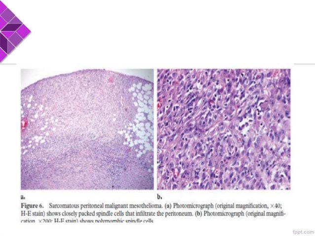
Sarcomatoid to an epithelioid appearance.

Mandala designs for coloring. Localized sarcomatoid mesotheliomas can only be diagnosed in the presence of spindle cell malignancies that exhibit immunoreactivity for cytokeratin and mesothelial markers and negative immunoreactivity for epithelial lesions in patients that show no multifocal or diffuse pleural spread and no evidence for extrapleural lesions. Immunohistochemically both the spindle shaped and epithelioid cells were at least focally positive for pancytokeratin vimentin calretinin a sma and desmin. The best discriminators among the antibodies considered to be negative markers for mesothelioma are cea moc 31 ber ep4 bg 8 and b723.
It is also found in certain types of lung cancers and breast cancers. Immunohistochemistry has a more limited role in the diagnosis and distinction of sarcomatoid mesothelioma from other spindle cell neoplasms. The combination of a broad spectrum cytokeratin with calretinin combines both high sensitivity 77 for ae1ae3 with high specificity 100 for calretinin for sarcomatoid mesothelioma and can be diagnostically useful.
Pathologists use cytokeratin 56 to stain cancer tissue samples. Based on these findings the tumor was diagnosed as sarcomatoid mesothelioma. Thirtyone malignant sarcomatoid mesotheliomas were studied.
Cytokeratin 5 and 56 in diagnosing mesothelioma. Differentiating sarcomatoid mesothelioma from other pleural based spindle cell tumours by light microscopy can be challenging especially in a biopsy. In addition s100 protein laminin and collagen iv are usually positive in true adipose tissue and can help in distinguishing 90 arch pathol lab medvol 142 january 2018 malignant mesothelioma diagnosishusain et al.
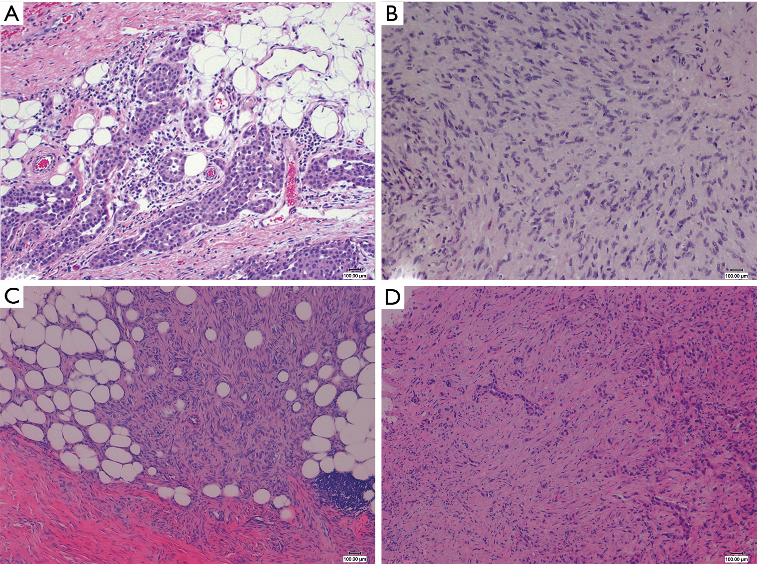
Regions horizontally oriented cytokeratin positive cells may be encountered around the fatlike spaces figure 6. Right scrotum and consisted of spindle shaped neoplastic cells that invaded the surrounding tissue. Cytokeratin 56 is a positive marker for malignant pleural mesothelioma found in more than three fourths of cases.
The role of immunohistochemistry in this differential diagnosis is not as well defined as it is for distinguishing epithelioid mesothelioma from adenocarcinoma. After analyzing the results it is concluded that calretinin cytokeratin 56 and wt1 are the best positive markers for differentiating epithelioid malignant mesothelioma from pulmonary adenocarcinoma.
More From Mandala Designs For Coloring
- Pusheen Colouring Pages Food
- Immigration Law Office
- Lol Pictures To Print And Colour
- Lake County Mesothelioma Attorney
- Mesothelioma Treatment National Cancer Institute
Incoming Search Terms:
- Cytokeratin Expression In Gastrointestinal Stromal Tumors Morphology Meaning And Mimicry Sing Y Ramdial Pk Ramburan A Sewram V Indian J Pathol Microbiol Mesothelioma Treatment National Cancer Institute,
- Https Patologi Com Guideline 20mesotheliom Pdf Mesothelioma Treatment National Cancer Institute,
- Pleomorphic Mesothelioma Report Of 10 Cases Modern Pathology Mesothelioma Treatment National Cancer Institute,
- Mesothelioma Lungs And Pleura Mypathologyreport Ca Ca Mesothelioma Treatment National Cancer Institute,
- The Pathological And Molecular Diagnosis Of Malignant Pleural Mesothelioma A Literature Review Ali Journal Of Thoracic Disease Mesothelioma Treatment National Cancer Institute,
- Https Encrypted Tbn0 Gstatic Com Images Q Tbn 3aand9gcqhjpudk4mltvqtva5hp2nrmoggfywdmzos5bx1le6l Sprigk Usqp Cau Mesothelioma Treatment National Cancer Institute,


