Pulmonary Mets Radiology, Https Www Atsjournals Org Doi Pdf 10 1513 Annalsats 201706 436cc
Pulmonary mets radiology Indeed recently is being hunted by users around us, maybe one of you personally. People now are accustomed to using the net in gadgets to view video and image information for inspiration, and according to the title of this article I will talk about about Pulmonary Mets Radiology.
- Grand Rounds Cannon Ball Secondaries And Pulmonary Metastasis Chest Medicine Made Easy Dr Deepu
- Pulmonary Metastases Colon Cancer Radiology Case Radiopaedia Org
- A Rare Case Of Giant Cell Tumour Gct Of Bone With Lung Metastases Bmj Case Reports
- Atypical Pulmonary Metastases Spectrum Of Radiologic Findings Radiographics
- Pulmonary Metastases Radiology Key
- Cannonball Lung Metastases As A Presenting Feature Of Ectopic Hcg Expression Sciencedirect
Find, Read, And Discover Pulmonary Mets Radiology, Such Us:
- Pulmonary Metastases Radiology Reference Article Radiopaedia Org
- Cavitating Lung Metastasis Secondary To Ameloblastoma Saheer S Enose P Thangakunam B Irodi A Korula A Lung India
- Bone Metastases Radiology Key
- 3
- Atypical Pulmonary Metastases Spectrum Of Radiologic Findings Radiographics
- Simple Easter Coloring Pages
- Veterans With Mesothelioma Facts And Figures
- Signs And Symptoms Of Asbestosis
- Mesothelioma Gross Specimen
- Lawsuit For Asbestos Exposure
If you are searching for Lawsuit For Asbestos Exposure you've come to the right place. We have 104 graphics about lawsuit for asbestos exposure including images, photos, photographs, wallpapers, and much more. In these web page, we additionally have variety of images available. Such as png, jpg, animated gifs, pic art, logo, black and white, translucent, etc.
Although not used routinely mri may be as sensitive in the detection of pulmonary metastases as ct 24.
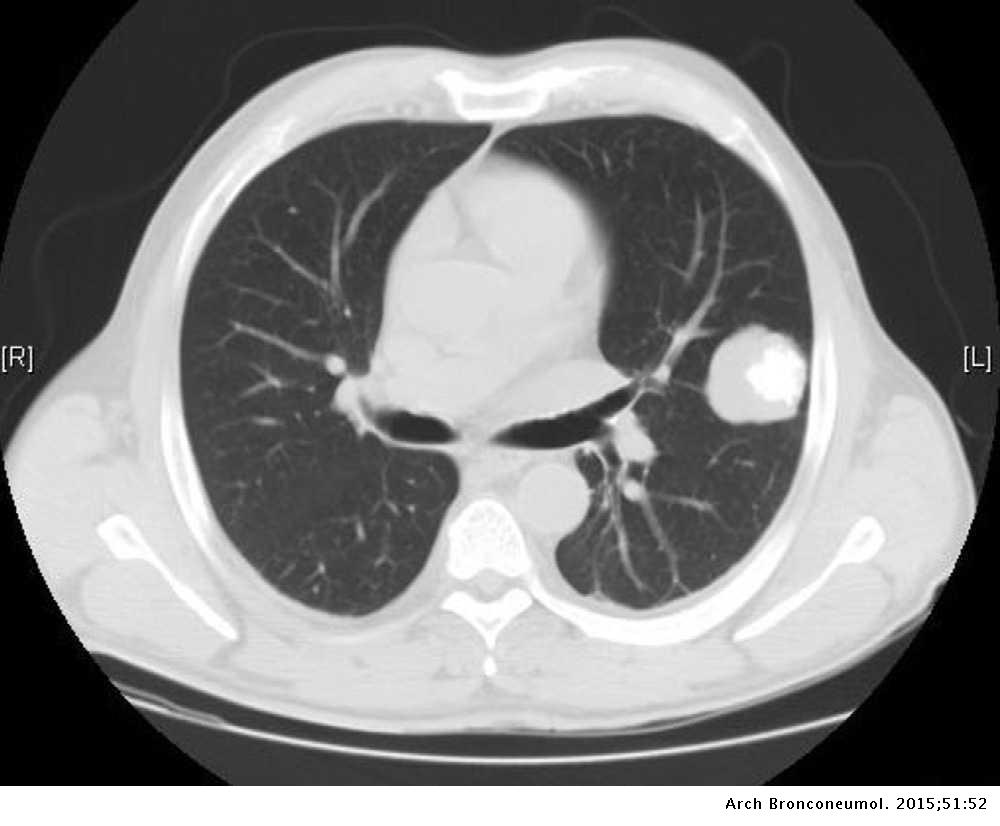
Lawsuit for asbestos exposure. Routine chest ct scans may reveal peripheral nodules as small as 2 3 mm and high resolution ct may demonstrate lymphangitic carcinomatosis. These should not be confused with metastatic pulmonary calcification. Cannonball metastases lungs cannonball metastases refer to multiple large well circumscribed round pulmonary metastases that appear not unsurprisingly like cannonballs.
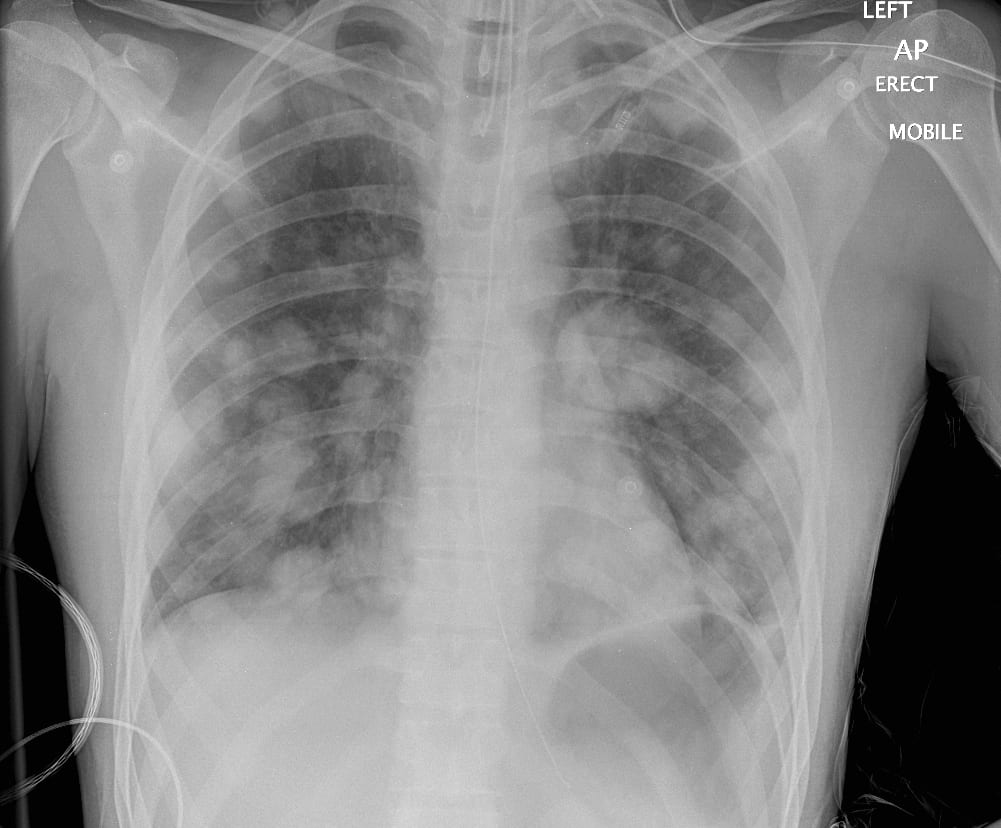
Transitional cell carcinoma of bladder 3. Typical radiologic findings of a pulmonary metastasis include multiple peripherally located round variable sized nodules hematogenous metastasis and diffuse thickening of the interstitium lymphangitic carcinomatosis 5 7. Computed tomography ct is clearly more sensitive than chest radiography or conventional linear tomography in the detection of pulmonary metastases.
Radiography lung nodules. The french terms envolee de ballons and lacher de ballons which translate to balloons release are also used to describe this same appearance. A halo of ground glass opacity representing hemorrhage can be seen particularly surrounding hemorrhagic pulmonary metastases such as choriocarcinoma and angiosarcoma 1.
Typical radiologic ndings of a pulmonary metastasis include multiple round variable sized nodules and diffuse thickening of interstitium. Metastases with such an. Cavitation is thought to occur in around 4 of lung metastases 2.
Pulmonary metastases may result in four main types of imaging manifestations. Pathology calcification in metastases can arise through a variety of mechanisms. Calcifying pulmonary metastases are rare.
Cavitary pulmonary metastases are most commonly 70 caused by squamous cell carcinoma which may of the lung or head and neck 146. In daily practice however unusual radiologic features of metastases are frequently encountered that make distinction from other nonmalignant pulmonary. Other primaries are varied and include.
The american college of radiology acr recommends that cxr should be the initial imaging modality used in the screening of pulmonary metastasis in patients with known extrathoracic malignancy. Dr bruno di muzio and dr yuranga weerakkody et al. Cystic pulmonary metastases are atypical morphological form on pulmonary metastases where lesions manifest as distinct cystic lesions.
The most common manifestation of pulmonary metastases consists of multiple nodules most numerous in the basal portions of the lungs reflecting the effect of gravity on blood flow. Among cases of multiple nodules detected with ct 73 were reported to be pulmonary metastases 8.
More From Lawsuit For Asbestos Exposure
- Pineapple Coloring Pages
- Mesothelioma Family Study
- Recent Mesothelioma Settlements Uk
- Mulan Coloring Pages
- Cincinnati Mesothelioma Law Firm Mesothelioma Lawyer
Incoming Search Terms:
- Right Upper Lobe Lung Cancer With Rib Metastasis Radiologypics Com Cincinnati Mesothelioma Law Firm Mesothelioma Lawyer,
- Lung Cancer With Bone Metastasis Radiology At St Vincent S University Hospital Cincinnati Mesothelioma Law Firm Mesothelioma Lawyer,
- Pathology Outlines Metastases Cincinnati Mesothelioma Law Firm Mesothelioma Lawyer,
- Cannonball Lung Metastases As A Presenting Feature Of Ectopic Hcg Expression Sciencedirect Cincinnati Mesothelioma Law Firm Mesothelioma Lawyer,
- Pulmonary Metastases Radiology Key Cincinnati Mesothelioma Law Firm Mesothelioma Lawyer,
- Two Synchronous Lung Metastases From Malignant Melanoma The Same Patient But Different Morphological Patterns Sciencedirect Cincinnati Mesothelioma Law Firm Mesothelioma Lawyer,


