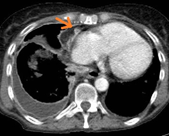
Pleural Thickening Ct Scan, Radiological Review Of Pleural Tumors
Pleural thickening ct scan Indeed recently has been sought by consumers around us, perhaps one of you. Individuals now are accustomed to using the internet in gadgets to see video and image information for inspiration, and according to the title of this post I will talk about about Pleural Thickening Ct Scan.
- Https Www Resmedjournal Com Article S0954 6111 17 30033 1 Pdf
- Pleura Thickening An Overview Sciencedirect Topics
- Pleural Thickening Radiology Reference Article Radiopaedia Org
- Chyliform Effusion Without Pleural Thickening In A Patient With Rheumatoid Arthritis A Case Report
- Chest Ct Scan Demonstrating Pleural Thickening Of The Inferior Half Of Download Scientific Diagram
- The Role Of 18f Fdg Pet Ct Integrated Imaging In Distinguishing Malignant From Benign Pleural Effusion
Find, Read, And Discover Pleural Thickening Ct Scan, Such Us:
- Pleural Thickening Radiology Reference Article Radiopaedia Org
- Https Journal Chestnet Org Article S0012 3692 18 30641 X Pdf
- Investigating Pleural Thickening The Bmj
- Epos Trade
- Pleural Malignant Mesothelioma With Micropapillary Pattern A Case Report And Literature Review
- Divorce Lawyers I Shaved My Head
- Unicorn Shoppies Coloring Pages
- Disney Animal Coloring Pages
- Mesothelioma Claim Amounts Uk
- Picnic Pops Coloring Pages
If you are looking for Picnic Pops Coloring Pages you've arrived at the perfect place. We ve got 104 images about picnic pops coloring pages adding pictures, pictures, photos, wallpapers, and much more. In such page, we also have variety of images out there. Such as png, jpg, animated gifs, pic art, logo, black and white, translucent, etc.
B composite image with coronal reformatted ct scans in soft tissue and lung windows shows the radiographic opacity is composed mostly of extrapleural fat arrows in addition to pleural thickening.

Picnic pops coloring pages. The undulating inferior margin of the apical cap is better seen in lung window. Ct is the work horse of pleural imaging able to achieve specificities of close to 100 3. According to etiology it may be classified as.
The apex of the lung was the most frequently affected area additional file 1. Ct scans can detect early signs of pleural thickening when scar tissue is 1 2 mm in thickness. Ct features suggestive of malignancy include circumferential pleural thickening sensitivity 41 specificity 100 parietal pleural thickening 1 cm 36 94 nodularity 51 94 and mediastinal pleural involvement 56 884 5 the british thoracic societys guidelines for the management of mesothelioma suggest consideration of a 60.
This is symmetric bilaterally and measures less than 5 mm in height. Pet and mri scans provide even further detail and may be used to differentiate pleural thickening from malignant mesothelioma. Could this cause pleural thickening.
Often pleural thickening is diagnosed alongside its cause. I have to have a ct scan in the next three weeks because of pleural thickening in the left lung. Pet and mri scans doctors can use positron emission tomography scans and magnetic resonance imaging scans to distinguish between pleural thickening and pleural mesothelioma a cancer affecting the pleura and often associated with asbestos exposure.
2amore than half of the cases were bilateral and 357 involved thickening on the. Highlighter august 6 2010 at anon85531 there is not much research on the effects of carbon particles on the lungs but i was able to find a source of information for you. Table s2pleural thickening involving the apical area of either lung was defined as an apical cap which accounted for 922 n 836907 of the cases fig.
A physical examination may also help diagnose the condition. Pleural thickening is often diagnosed with imaging scans such as computed tomography ct scans. The portal venous phase also known as the pleural phase in the chest is.
Pleural thickening was found predominantly at the apex of the right lung. The american journal of. It can occur with both benign and malignant pleural disease.
There was a faint opacity in both lungs sinus costo phrenicus of left lung was blunt and the pleural thickening existed. An x ray can reveal thickening of the pleura but a ct scan is more likely to show thickening in better detail. In cases where multiple nodular regions or pleural thickening are present the diagnosis may be evident especially if a primary tumor or other metastatic deposits are visible.
A ct scan which is also used to diagnose asbestosis and pleural plaques can confirm the condition earlier. In chest ct scan there was pleural thickening in the posterobasal of the chest wall and dissemina ting to the mediastinum paraaortic figure 2 figure 1.
More From Picnic Pops Coloring Pages
- Mesothelioma Treatment Affected Lung
- Gpwlaw Wv West Virginia Mesothelioma Lawyer
- Kendall Jenner Nba Lineup
- Mrs Meyers Surface Cleaner
- Frozen Coloring Pages Pdf
Incoming Search Terms:
- Covid 19 Pneumonia Siemens Healthineers Danmark Frozen Coloring Pages Pdf,
- Radiological Review Of Pleural Tumors Frozen Coloring Pages Pdf,
- Https Encrypted Tbn0 Gstatic Com Images Q Tbn 3aand9gct5wagysvxv6irl7dgfam2 Tlmenkhg96a8naqacd 7ymwnujus Usqp Cau Frozen Coloring Pages Pdf,
- Fibrothorax With Pleural Thickening Radiology Case Radiopaedia Org Frozen Coloring Pages Pdf,
- Https Www Resmedjournal Com Article S0954 6111 17 30033 1 Pdf Frozen Coloring Pages Pdf,
- View Image Frozen Coloring Pages Pdf,


