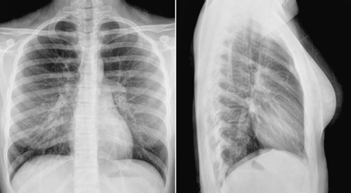
Peripheral Lung Markings, Https Encrypted Tbn0 Gstatic Com Images Q Tbn 3aand9gcthebtbnqbojdnknihzobt6 Qvai 4jlnrfclapzpwpwx Iapld Usqp Cau
Peripheral lung markings Indeed lately has been sought by users around us, perhaps one of you. Individuals now are accustomed to using the internet in gadgets to see video and image data for inspiration, and according to the title of the post I will discuss about Peripheral Lung Markings.
- Https Www Bir Org Uk Media 258608 Mark Rodriguez Philips Trainee For Excellence Unofficial Guide To Radiology Pdf
- Linear Lung Density X Ray Dr Ahmed Esawy
- Chest Medicine Made Easy Dr Deepu
- The Role Of Chest Radiography In Confirming Covid 19 Pneumonia The Bmj
- Pulmonary Embolism Chest X Ray Signs Epomedicine
- Pulmonary Vascularity Radiology Key
Find, Read, And Discover Peripheral Lung Markings, Such Us:
- Chest X Ray Showing Decreased Peripheral Lung Vascular Markings And Download Scientific Diagram
- Https Www Ajol Info Index Php Cme Article View 43826 27345
- Chest Radiograph Showing Increased Lung Markings In The Right Lower Download Scientific Diagram
- The Radiology Assistant Lung Disease
- Interstitial Changes Chest X Ray Medschool
- Free Easter Bunny Coloring Pages
- Mesothelioma And Asbestos Cancer
- Riverbank State Park Ice Rink
- Pumpkin Carving Ideas Printable
- Little Pumpkin Faces
If you are looking for Little Pumpkin Faces you've arrived at the perfect place. We have 104 images about little pumpkin faces adding images, photos, photographs, wallpapers, and much more. In these web page, we additionally have variety of graphics out there. Such as png, jpg, animated gifs, pic art, symbol, black and white, transparent, etc.
Emphysema with increased pulmonary markings.

Little pumpkin faces. The margins of the enlarged vessels are sharp and. Panlobular emphysema is diffuse and is most severe in the lower lobes. Infarction peripheral consolidation in a patient with acute shortness of breath with low oxygen level and high d dimer.
It can either mean a plain film or hrctct feature. Bronchiectasis asthma and most often seen in interstitial pulmonary edema. Organizing pneumonia op multiple chronic consolidations.
4 prominent pulmonary vessels of an irregular and indistinct contour and often with cor pulmonale. Its a subjective call by the reading physician regarding the prominence of the connective tissue in the lung. Pulmonary vessels in the affected lung appear fewer and smaller than normal.
In severe panlobular emphysema the characteristic appearance of extensive lung destruction and the associated paucity of vascular markings are easily distinguishable from normal lung parenchyma. The apical parts of the lungs are devoid of sizable vascular markings in most normal individuals. 3 rapid peripheral tapering of pulmonary vessels and their unequal distribution often with the presence of bullae.
Reticular interstitial pattern is one of the patterns of linear opacification in the lung. Emphysema with decreased pulmonary markings. Although the pulmonary vessels show some asymmetric anatomy in the lung hila the pulmonary vascularity in the lungs is normally symmetric.
Pulmonary hemorrhage in a patient with hemoptoe. The peripheral pulmonary arteries can be tortuous. It may also be called a spot on the lung or a coin lesion pulmonary nodules are smaller than three centimeters around 12 inches in diameter.
Pathology causes reticulation can be subdivided by the size of the intervening pulmonary lucency in. If the growth is larger than that it is called a pulmonary mass and is more likely to represent a cancer than a nodule. On a radiograph interstitial lung markings are fine white lines and dots lines seen end on that represent the pulmonary interstitium.
Subtle edema excess amount of fluid in the lungs from heart weakness kidney dysfunction ot. What does that mean answered by dr. A pulmonary nodule is a small round or oval shaped growth in the lung.
Chest ex ray says lungs show increased interstitial markings with no active infiltratemass or effusion. Increased markings can be. Thickening of the peripheral interstitium either medially or laterally produces kerley lines axial interstitial thickening is difficult to distinguish from airways disease that result in bronchial wall thickening eg.
More From Little Pumpkin Faces
- What Causes Mesothelioma
- Lol Pet Coloring Pages
- Malignant Mesothelioma Pleural Markers
- Mesothelioma Childhood Exposure
- Red Sox Coloring Pages
Incoming Search Terms:
- Im Tutor Msu Radiology Department Red Sox Coloring Pages,
- B9w7 L7 Pulmonary Htn And Embolic Diseases Respiratory Flashcards Memorang Red Sox Coloring Pages,
- Chest Radiology Red Sox Coloring Pages,
- Https Www Bir Org Uk Media 258608 Mark Rodriguez Philips Trainee For Excellence Unofficial Guide To Radiology Pdf Red Sox Coloring Pages,
- Lagcnyp10t 2pm Red Sox Coloring Pages,
- Reading Chest X Rays Updated The Book Of Oi Red Sox Coloring Pages,


