Peripheral Lung Cancer Radiology, Ct Features Of Lung Scar Cancer Gao Journal Of Thoracic Disease
Peripheral lung cancer radiology Indeed recently is being sought by consumers around us, maybe one of you personally. Individuals now are accustomed to using the internet in gadgets to view video and image data for inspiration, and according to the name of this article I will talk about about Peripheral Lung Cancer Radiology.
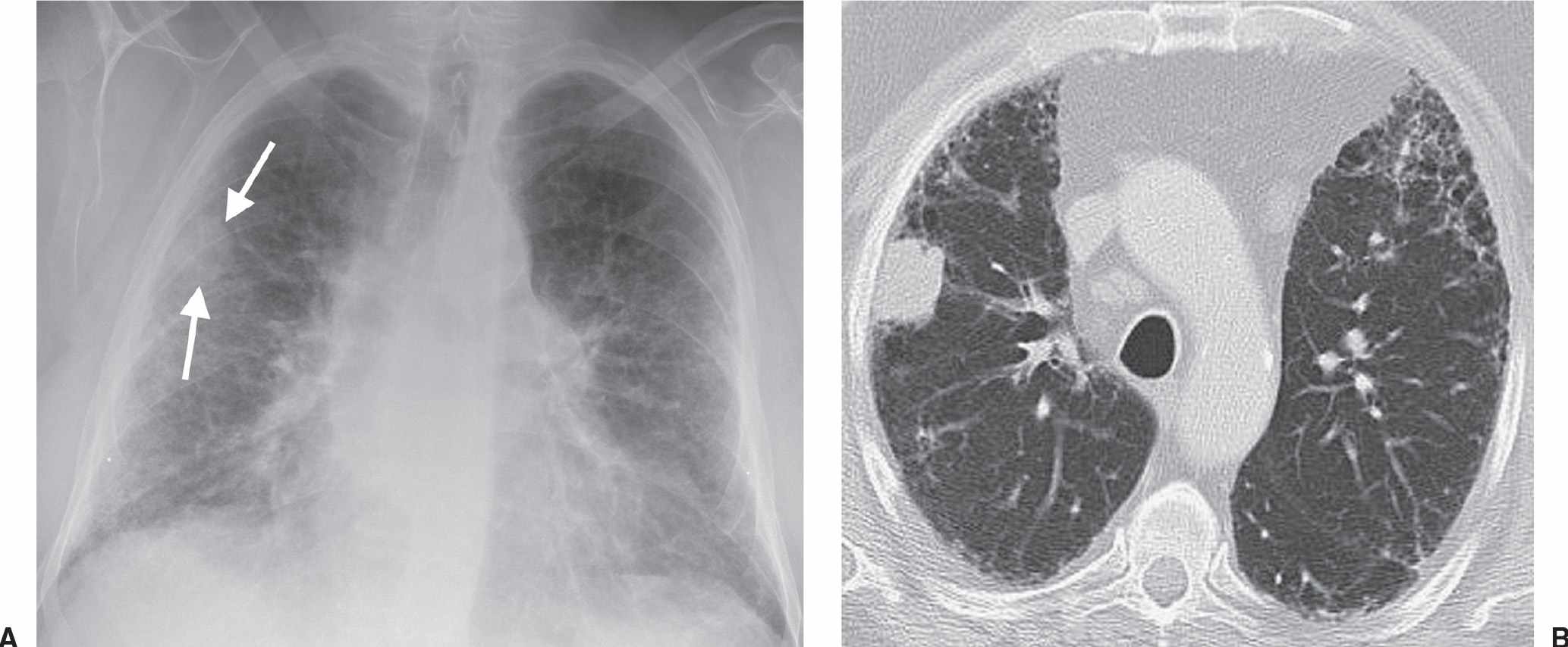
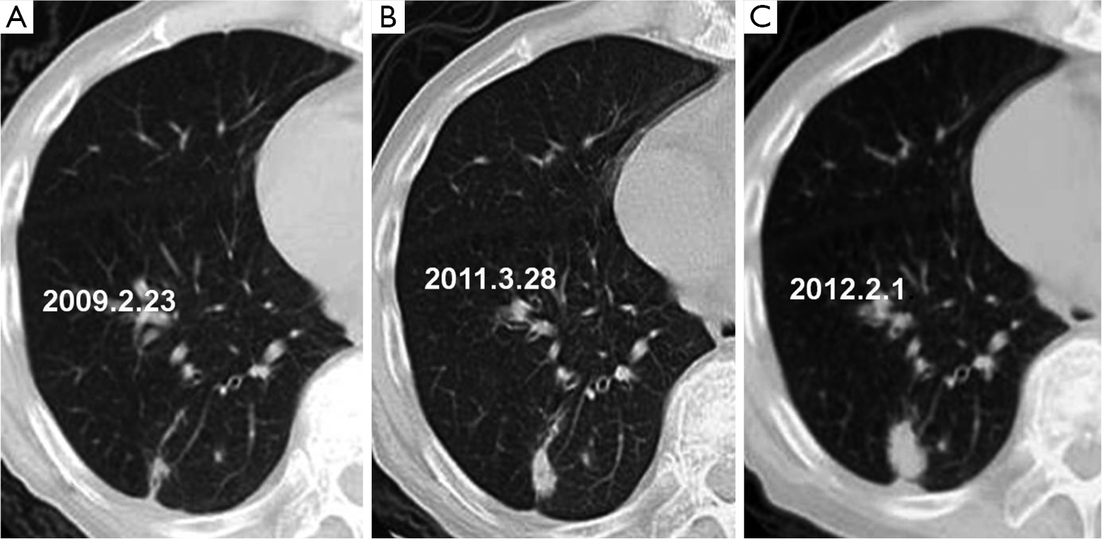
- Ct Features Of Lung Scar Cancer Gao Journal Of Thoracic Disease
- Plos One Detection Of Rib Metastases In Patients With Lung Cancer A Comparative Study Of Mri Ct And Bone Scintigraphy
- Pathology Outlines Metastases
- Ground Glass Opacity Lung Nodules In The Era Of Lung Cancer Ct Screening Radiology Pathology And Clinical Management Cancer Network
- Computer Aided Detection Of Peripheral Lung Cancers Missed At Ct Roc Analyses Without And With Localization Radiology
- Peripheral Intrapulmonary Lymph Node Metastases Of Non Small Cell Lung Cancer The Annals Of Thoracic Surgery
Find, Read, And Discover Peripheral Lung Cancer Radiology, Such Us:
- Cavitating Lung Cancer Radiology Case Radiopaedia Org
- Differential Diagnosis Of Solitary Pulmonary Inflammatory Lesions And Peripheral Lung Cancers With Contrast Enhanced Computed Tomography
- Clinmed International Library The Many Faces Of Lung Cancer International Journal Of Cancer And Clinical Research
- What Clinician S Need To Know About Imaging Features In Lung Cancer Sureka B Mittal Mk Mittal A Sinha M Thukral Bb J Mahatma Gandhi Inst Med Sci
- Neoplasms Of The Lung Radiology Key
- Otto Hasibuan Law Firm
- Non Asbestos Related Mesothelioma
- Pocoyo Coloring Pages Printable
- Mesothelioma Treatment Attorney Advertising
- What Is Mesothelioma And What Causes It
If you re searching for What Is Mesothelioma And What Causes It you've arrived at the ideal location. We ve got 104 graphics about what is mesothelioma and what causes it including images, pictures, photos, backgrounds, and more. In such web page, we also provide number of graphics available. Such as png, jpg, animated gifs, pic art, logo, blackandwhite, translucent, etc.
Most peripheral carcinoid tumors tend to involve a subsegmental bronchus 2.
What is mesothelioma and what causes it. Evolution of peripheral lung adenocarcinomas. Imaging features are often non specific and tissue diagnosis is essential in determining diagnosis. Low dose spiral ct parameters were 120 kvp 50 ma 10 mm collimation and 21 pitch.
Ct appearance growth rate location and histologic features of 61 lung cancers. The disease is very common and in its earliest stages 70 of cases can be cured by surgery 4despite this lung cancer has an overall prognosis so dismal that incidence exceeds prevalence 5the main risk factor smoking is easily identifiable and noninvasive screening tests such as chest radiography and sputum cytology are widely. The areas of focus include image based evaluation of tumor growth rates and tumor burden kinetics radiomics and molecular and functional imaging.
13 aoki t nakata h watanabe h et al. In response to the recent advances of precision lung cancer therapy imaging strategies of treatment monitoring have also been evolving. Many have a lobulated margin with an average hounsfield value on postcontrast imaging of 50 2.
Consolidation any pathologic process that fills the alveoli with fluid pus blood cells including tumor cells or other substances resulting in lobar diffuse or multifocal ill defined opacities. To evaluate the prognostic importance of thin section computed tomographic ct findings of peripheral lung adenocarcinomas. Emerging strategies of precision imaging for lung cancer.
Primaries that metastasize as endobronchial deposits can include. Ct findings correlated with histology and tumor doubling time. This article will broadly discuss all the histological subtypes as a group focusing on their common aspects.
Five year lung cancer screening experience. Interstitial involvement of the supporting tissue of the lung parenchyma. Lung cancer primary lung cancer or frequently if somewhat incorrectly known as bronchogenic carcinoma is a broad term referring to the main histological subtypes of primary lung malignancies that are mainly linked with inhaled carcinogens with cigarette smoke being a key culprit.
May have a sensitivity of around 75 7. The subjects were 127 patients with adenocarcinomas smaller than 3 cm in largest diameter who underwent at least a lobectomy with hilar and mediastinal lymphadenectomy. The margin characteristics of nodules and the extent of ground glass.
Recognize a pattern of peripheral lung disease on chest radiography or computed tomography ct and give an appropriate differential diagnosis including a single most likely diagnosis when supported by associated radiologic findings or clinical information eg peripheral lung disease associated with paratracheal and bilateral hilar lymphadenopathy in an asymptomatic. Peripheral lung cancer was detected in 15 of 3457 examinations 03. Pulmonary metastases typically appear as peripheral rounded nodules of variable size scattered throughout both lungs 1.

Clinmed International Library The Many Faces Of Lung Cancer International Journal Of Cancer And Clinical Research What Is Mesothelioma And What Causes It
More From What Is Mesothelioma And What Causes It
- Mesothelioma Radiation Rusch
- Legal Aid Ontario Find A Lawyer
- Modern Law Firm Sign
- Fish Coloring Pages For Kids
- Long Term Exposure To Asbestos
Incoming Search Terms:
- Lung Cancer Radiology Reference Article Radiopaedia Org Long Term Exposure To Asbestos,
- Neoplasms Of The Lung Radiology Key Long Term Exposure To Asbestos,
- Lung Cancers Flashcards Memorang Long Term Exposure To Asbestos,
- Peripheral Intrapulmonary Lymph Node Metastases Of Non Small Cell Lung Cancer The Annals Of Thoracic Surgery Long Term Exposure To Asbestos,
- Https Www Annalsthoracicsurgery Org Article S0003 4975 02 03895 X Pdf Long Term Exposure To Asbestos,
- X Ray Imaging For Covid 19 Patients Long Term Exposure To Asbestos,


