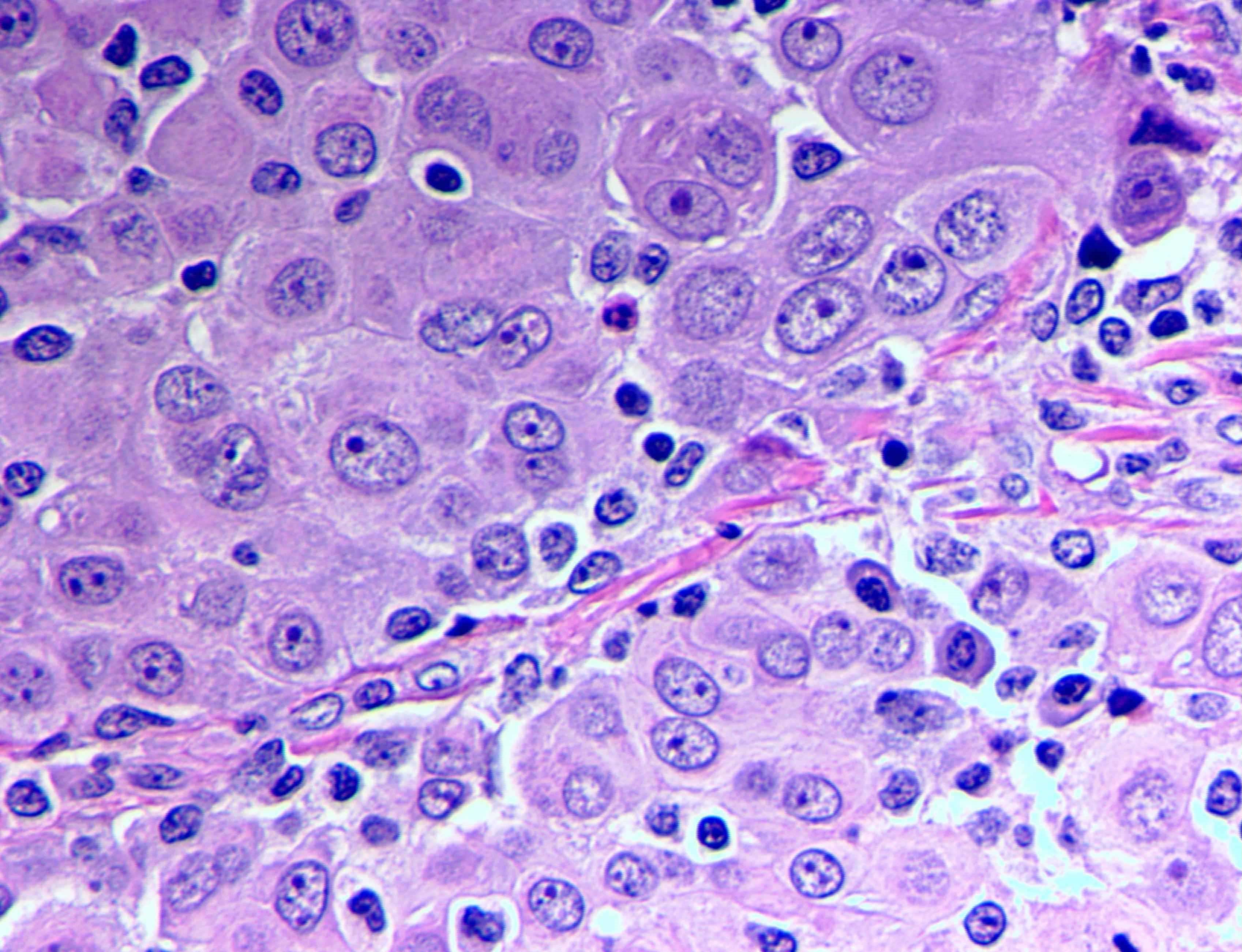
Most Common Histopathology Of Mesothelioma, Frontiers New Perspectives On Diagnosis And Therapy Of Malignant Pleural Mesothelioma Oncology
Most common histopathology of mesothelioma Indeed lately is being hunted by users around us, perhaps one of you. Individuals now are accustomed to using the internet in gadgets to see video and image information for inspiration, and according to the title of the post I will talk about about Most Common Histopathology Of Mesothelioma.
- 2016 Evening Specialty Conference Gynecologic Pathology
- Molecular Characterization Of Localized Pleural Mesothelioma Modern Pathology
- Mesothelioma Blog Mesothelioma Pathology Dermoid Cyst Rare Genetic Disorders
- Malignant Peritoneal Mesothelioma In A Patient With Intestinal Fistula Incisional Hernia And Abdominal Infection A Case Report
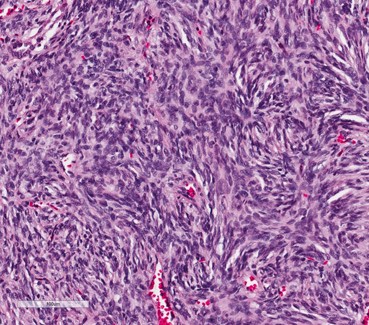
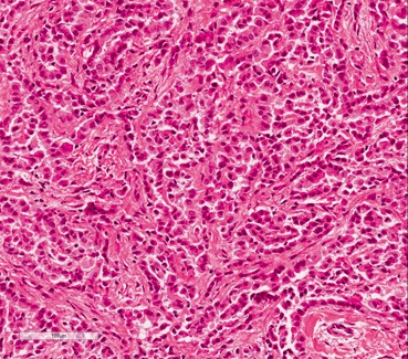
- Mesothelioma Cell Types Epithelioid Sarcomatoid And Biphasic Cells
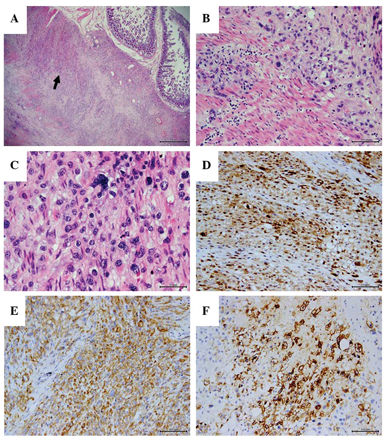
- Metastasis Of Mesothelioma To The Maxillary Gingiva
Find, Read, And Discover Most Common Histopathology Of Mesothelioma, Such Us:
- Malignant Mesothelioma And Other Mesothelial Proliferations Chapter 28 Modern Soft Tissue Pathology
- Metastasis Of Mesothelioma To The Maxillary Gingiva
- Fatal Hemothorax Caused By Pleural Mesothelioma In A Lion
- Https Academic Oup Com Ajcp Article Pdf 110 3 397 24884795 Ajcpath110 0397 Pdf
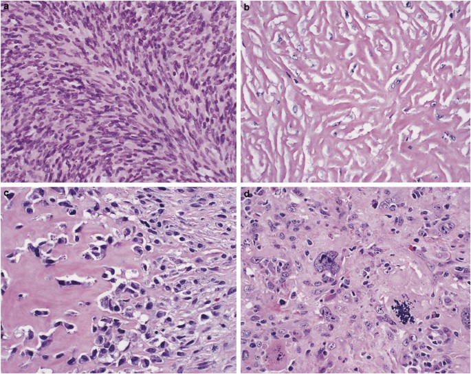
- Sarcomatoid Mesothelioma A Clinical Pathologic Correlation Of 326 Cases Modern Pathology
- Equestria Coloring Pages
- How Can Mesothelioma Affect Your Body
- Free Printable Thanksgiving Coloring Sheets
- World Best Lawyers 2020
- Mesothelial Cells In Pleural Fluid Cytology
If you are searching for Mesothelial Cells In Pleural Fluid Cytology you've come to the perfect location. We have 100 graphics about mesothelial cells in pleural fluid cytology including images, pictures, photos, wallpapers, and more. In such page, we additionally provide number of graphics available. Such as png, jpg, animated gifs, pic art, logo, blackandwhite, translucent, etc.
Imaging scans are often used to properly identify these rarer cells.

Mesothelial cells in pleural fluid cytology. The researchers compared the presence of several markers eg pan cytokeratin cytokeratin 56 wt 1 calretinin and thrombomodulin in the three types of cancer. Epithelioid cells are well defined with a single nucleus and tend to clump. Signs and symptoms of mesothelioma may.
There is a well established link between mesothelioma and a sbestos. The most common mesothelioma cell type epithelioid cells are recognized by cytologists because of their uniform and defined appearance. A 2003 study in histopathology analyzed the makeup of some of sarcomatoid mesotheliomas most common mimics high grade sarcoma and pulmonary sarcomatoid carcinoma.
Less commonly the lining of the abdomen and rarely the sac surrounding the heart or the sac surrounding the testis may be affected. These rare mesothelioma cell subtypes require additional care to best understand how theyre reacting in order to most effectively treat the cancer. Mesothelioma is a malignant tumour arises from mesothelial lining of pleura peritoneum pericardium and tunica vaginalis pleural mesothelioma is the most common of these.
The most common area affected is the lining of the lungs and chest wall. This process is part of mesothelioma pathology which involves examining either tissue or fluid to determine if this cancer exists in the body. They contain a single nucleus and tend to grow slower than other cancerous cell types.
Mesothelioma histology or mesothelioma histopathology is the study of tissue for the presence of mesothelioma. This is a good thing considering that epithelioid mesothelioma is the easiest of the types to treat and typically coincides with the best prognosis. Histopathology becomes even more important when the cells do not fall neatly into one of the three main mesothelioma cell types.
While epithelial cells multiply quickly they have a tendency to lump together which makes their overall tumor growth. Defined by single layer of surface mesothelial cells that lost bap1 expression usually presenting as unilateral pleural effusion no evidence of tumor by imaging or by direct examination of pleura no invasive mesothelioma developing for at least 1 year. Malignant mesothelioma in situ histopathology 2018721033.
Mesothelioma is a type of cancer that develops from the thin layer of tissue that covers many of the internal organs known as the mesothelium.
More From Mesothelial Cells In Pleural Fluid Cytology
- Is Mesothelioma
- Left Knee Malignant Mesothelioma
- Clark Hill Law
- Sugar Skull Coloring Pages
- Inside Out Coloring Pages Pdf
Incoming Search Terms:
- Histopathology Images Of Malignant Mesothelioma By Pathpedia Com Pathology E Atlas Inside Out Coloring Pages Pdf,
- Mesothelioma Cell Types Epithelioid Sarcomatoid And Biphasic Cells Inside Out Coloring Pages Pdf,
- 2016 Evening Specialty Conference Gynecologic Pathology Inside Out Coloring Pages Pdf,
- Ijms Free Full Text Heterogeneity In Malignant Pleural Mesothelioma Html Inside Out Coloring Pages Pdf,
- Pathology Outlines Mesothelioma Epithelioid Inside Out Coloring Pages Pdf,
- Pathology Outlines Diffuse Malignant Mesothelioma Inside Out Coloring Pages Pdf,
