Mesothelium Histology, Mesothelium Wikiwand
Mesothelium histology Indeed lately is being hunted by users around us, perhaps one of you. Individuals are now accustomed to using the internet in gadgets to see image and video data for inspiration, and according to the title of this article I will talk about about Mesothelium Histology.
- Siu Som Histology Gi
- Histology And Immunohistochemistry Of Lesions In Mextag Mice Following Download Scientific Diagram
- Epithelial Tissue Histology Flashcards Memorang
- Histology Quiz 1 Identification Slides L Simple Squamous Epithelium Mesothelium R Simple Squamous Epithelium Ppt Download
- Normal Histology
- Histology Virtual Lab Epithelial Tissues
Find, Read, And Discover Mesothelium Histology, Such Us:
- The Heart
- Http Staff Ui Ac Id System Files Users Jeanne Adiwinata Material Manual Inter Modul Cerna Pdf
- Description
- Mesothelium An Overview Sciencedirect Topics
- Mesothelioma Histology A Study Of Mesothelioma Cells
- Mickey Mouse Colouring Pages
- Coloring Pages 4 Kids
- Kilpatrick Law Firm
- Denise Cooney
- Chrysotile As A Cause Of Mesothelioma
If you re looking for Chrysotile As A Cause Of Mesothelioma you've come to the perfect place. We have 104 graphics about chrysotile as a cause of mesothelioma adding images, pictures, photos, wallpapers, and much more. In such page, we additionally have number of images out there. Such as png, jpg, animated gifs, pic art, logo, blackandwhite, translucent, etc.

Histology On Twitter The Arrow Points To The Mesothelium Which Is Part Of The Visceral Pleura Chrysotile As A Cause Of Mesothelioma
Histopathology is the study of diseased cells.
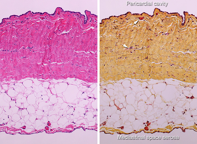
Chrysotile as a cause of mesothelioma. Peritoneal mesothelium produces peritoneal fluid. The mesothelium is a simple squamous epithelium that forms the lining of several body cavities pleural peritoneal pericardial cavities etc. While the mesothelium can aid in controlling tumors it is also.
Serosal mesothelium actively participates in various pathological. Mesothelium covers the outside surface of organs that project into the body cavities and thus often covers the normally convex surface of an organ. Although both of these are simple squamous epithelia the endothelial cells tend to have minimal cytoplasm such that there is little sign of cytoplasm between the bulging nuclei while the.
Mesothelium histology monolayer analyzing the ultrastructure connected with different mesothelial cells shows features which are different in squamous and also cuboidal type mesothelium. Mesothelial tissue also surrounds the male internal reproductive organs the tunica vaginalis testis and covers the internal reproductive organs of. The mesothelial cells are supported by a basal membrane in continuity with a thin layer of richly vascularized loose connective tissue with lymphatics and fatty tissue which is called the subserosa.
However immunostains can demonstrate invasion into underlying tissues differential diagnosis. The mesothelium is an important structure lining the chest abdomen and pelvis and serves not only to lubricate movements of organs in these regions but has important functions in fluid transport blood clotting and in resistance to infections and the spread of cancers. This process is part of mesothelioma pathology which involves examining either tissue or fluid to determine if this cancer exists in the body.
Histology guide a virtual histology laboratory with zoomable images of microscope slides and electron micrographs. The pleura thoracic cavity peritoneum abdominal cavity including the mesentery mediastinum and pericardium heart sac. Your pathologist will use histology techniques to provide the most accurate information about your mesothelioma cell type.
Histology is a branch of biology that involves the study of cells and tissues. Mesothelioma histology or mesothelioma histopathology is the study of tissue for the presence of mesothelioma. The mesothelium is a membrane composed of simple squamous epithelium that forms the lining of several body cavities.
Immunostains do not differentiate benign and malignant mesothelial cells as both are positive for keratin.
More From Chrysotile As A Cause Of Mesothelioma
- Different Kinds Of Asbestos
- What Does Asbestos Feel Like
- My Family Coloring Pages For Preschoolers
- Harry Potter Christmas Coloring Pages
- Fox Sports Radio Nyc
Incoming Search Terms:
- Mesothelial Cytopathology Libre Pathology Fox Sports Radio Nyc,
- Simple Squamous Epithelium Endothelium Endocardium Mesothelium Youtube Fox Sports Radio Nyc,
- Siu Som Histology Intro Fox Sports Radio Nyc,
- Unit5 Slide 14 Large Image Format Fox Sports Radio Nyc,
- Https Encrypted Tbn0 Gstatic Com Images Q Tbn 3aand9gcrlg3qse Aa8ft Za5qvj Lskzlxvhik5nfand Ikozotr58joe Usqp Cau Fox Sports Radio Nyc,
- Pin On Https Drawittoknowit Com Fox Sports Radio Nyc,


:background_color(FFFFFF):format(jpeg)/images/article/en/liver-histology/YgpzSzQwAuNMn8E0R9Jb0A_1AhNn1NNg34e7rDthjuUA_Liver.png)
