Mesothelial Cells Lung, Multiplexed Single Cell Transcriptomic Analysis Of Normal And Impaired Lung Development In The Mouse Biorxiv
Mesothelial cells lung Indeed lately has been hunted by consumers around us, perhaps one of you personally. People now are accustomed to using the net in gadgets to see image and video data for inspiration, and according to the name of this article I will discuss about Mesothelial Cells Lung.
- Mesothelial Hyperplasia An Overview Sciencedirect Topics
- Frontiers Free Floating Mesothelial Cells In Pleural Fluid After Lung Surgery Medicine
- Diverse Properties Of The Mesothelial Cells In Health And Disease In Pleura And Peritoneum Volume 1 Issue 2 2016
- Cell Types And Cellular Processes Involved In Lung Cancer Lc And Download Scientific Diagram
- Use Of Panel Of Markers In Serous Effusion To Distinguish Reactive Mesothelial Cells From Adenocarcinoma Subbarayan D Bhattacharya J Rani P Khuraijam B Jain S J Cytol
- Derivation Of Lung Mesenchymal Lineages From The Fetal Mesothelium Requires Hedgehog Signaling For Mesothelial Cell Entry Development
Find, Read, And Discover Mesothelial Cells Lung, Such Us:
- Derivation Of Lung Mesenchymal Lineages From The Fetal Mesothelium Requires Hedgehog Signaling For Mesothelial Cell Entry Development
- What Are Mesothelial Cells Mesotheliomahope Com
- Clinical Aspects Of Pleural Fluid Ph
- Lrrn4 And Upk3b Are Markers Of Primary Mesothelial Cells
- Clinicopathological Significance Of Microrna 21 In Extracellular Vesicles Of Pleural Lavage Fluid Of Lung Adenocarcinoma And Its Functions Inducing The Mesothelial To Mesenchymal Transition Watabe 2020 Cancer Medicine Wiley Online Library
- Mesothelioma Second Opinions
- Outline Pictures For Colouring Kindergarten
- Sight Word Coloring Pages Kindergarten
- Mesothelioma Support Uk
- Recharge Ashaway
If you are searching for Recharge Ashaway you've come to the ideal place. We ve got 104 graphics about recharge ashaway including pictures, photos, pictures, backgrounds, and more. In these webpage, we additionally have variety of images available. Such as png, jpg, animated gifs, pic art, symbol, blackandwhite, translucent, etc.
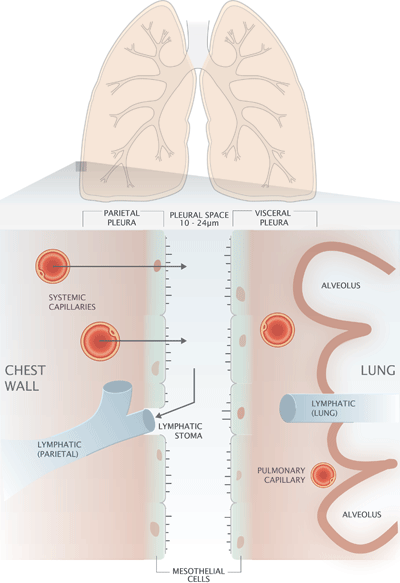
Some cells known as mesothelial cells line the pleura the thin double layered lining that covered the chest wall diaphragm and lungs.
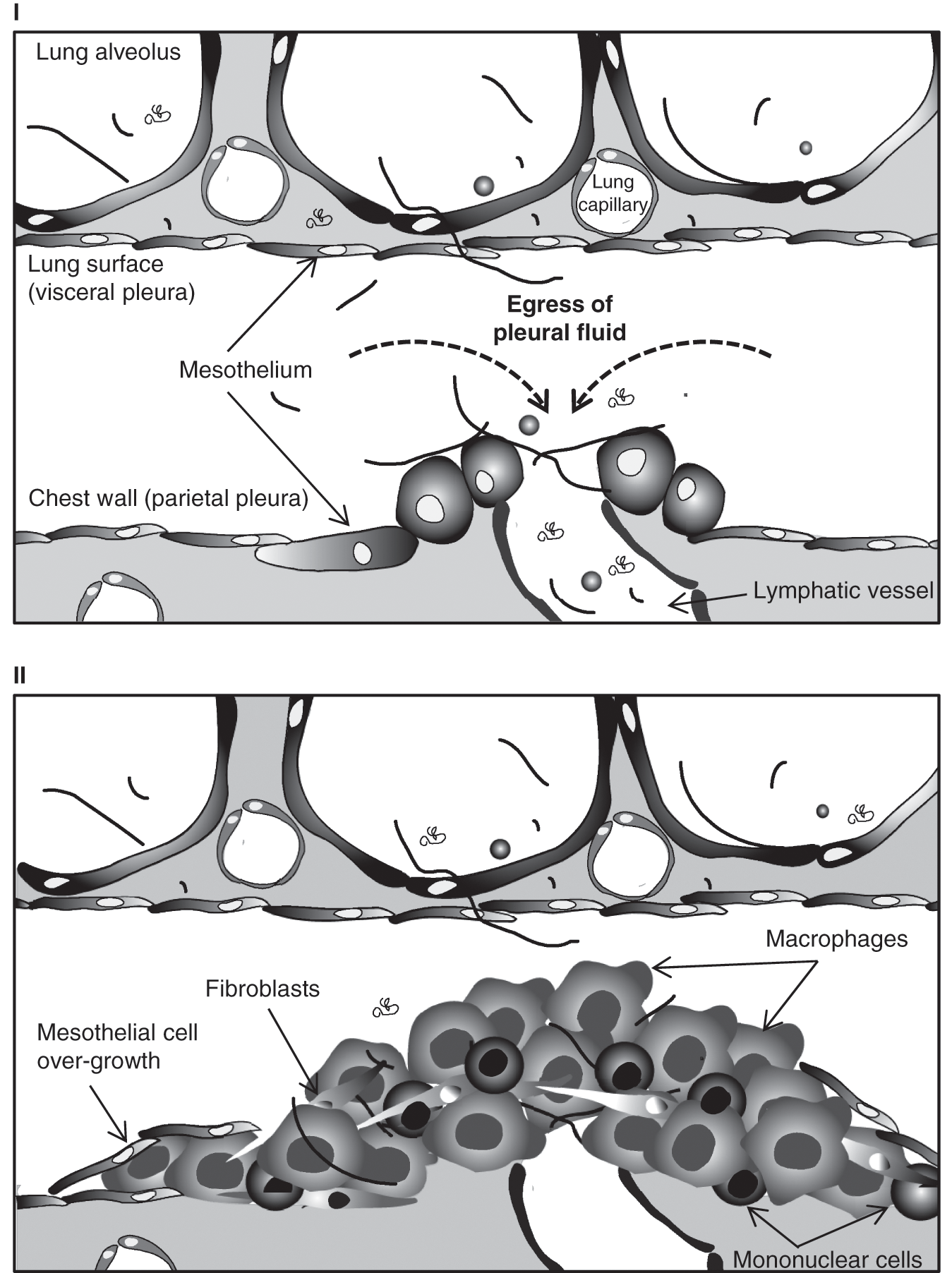
Recharge ashaway. There are certain cells that line the pleura the thin double layered lining which covers the lungs chest wall and diaphragm which are known as mesothelial cells. In this study our in vivo study revealed that pleural mesothelial cells pmcs migrated into lung parenchyma and localized alongside lung fibroblasts in sub pleural area in mouse pulmonary fibrosis. Pleural mesothelial cells pmc are metabolically dynamic cells that cover the lung and chest wall as a monolayer and are in intimate proximity to the underlying lung parenchyma.
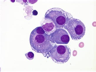
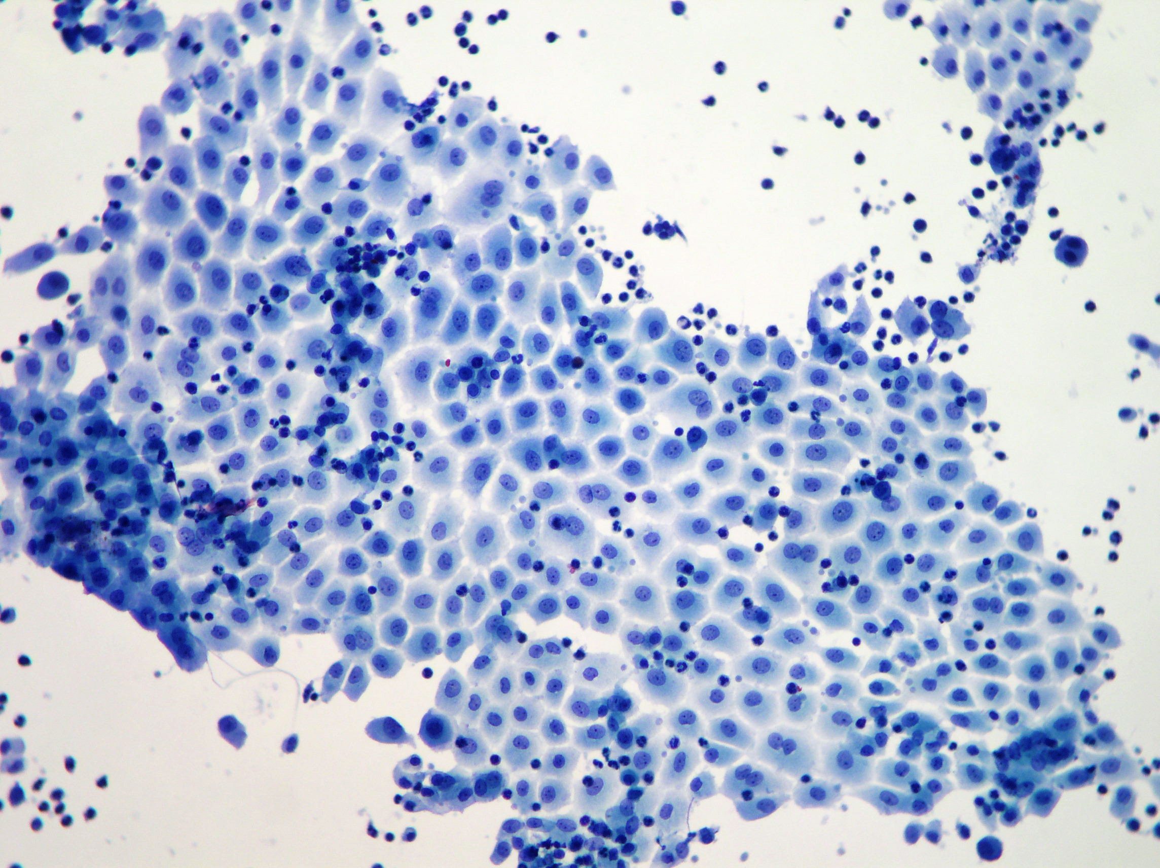
Mesothelial cells often appear like squamous cells when microscopically examined however despite their structure that resembles the squamous cells they are a unique form of an epithelial cell. Pleural mesothelial cells in pleural and lung diseases article literature review pdf available in journal of thoracic disease 76964 80 june 2015 with 120 reads how we measure reads. The origin of the myofibroblast in fibrotic lung disease is uncertain and no effective medical therapy for fibrosis exists.
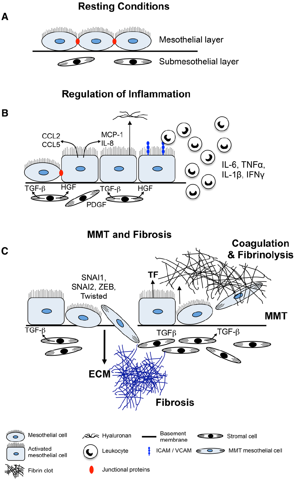
The mesothelium is a single continuous layer of epithelial cells that is divided into three primary regions. We have previously demonstrated that transforming growth factor b1 tgf b1 induces pleural mesothelial cell pmc transformation into myofibroblasts and haptotactic migration in vitrowhether pmc differentiation and migration occurs in vivo and whether this response. Pleural mesothelial cells pmcs undergo a process called mesothelial mesenchymal transition mesomt by which pmcs acquire a profibrotic phenotype characterized by cellular enlargement and elongation increased expression of a smooth muscle actin a sma and matrix proteins including collagen 1.
Mesothelial cells begin as the mesoderm during development the lungs derive from endoderm and apparently play an important part in the development of the lung. In the lung mesothelioma cells express high levels of cd44 and conditioned medium from mesothelioma cell lines stimulates hyaluronan synthesis in mesothelial cells. Markedly increased numbers of.
Our in vitro study displayed that cultured pmcs medium induced lung fibroblasts transforming into myofibroblast cultured fibroblasts medium promoted mesothelial mesenchymal transition of pmcs. When mesothelial cells are examined under the microscope they often appear like squamous cells. The precise role of pmc in the pathogenesis of pulmonary parenchymal fibrosis remains to be identified.
Mesothelial cells are found in variable numbers in most effusions but their presence at greater than 5 of total nucleated cells makes a diagnosis of tb less likely.
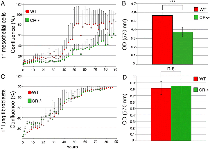
Overexpression Or Absence Of Calretinin In Mouse Primary Mesothelial Cells Inversely Affects Proliferation And Cell Migration Respiratory Research Full Text Recharge Ashaway
More From Recharge Ashaway
- Lydia Beetlejuice Costume
- Payout For Mesothelioma Victims Uk
- Show Pictures Of Zombies
- Postmortem Mesothelioma Diagnosis
- Up Coloring Book
Incoming Search Terms:
- Mesothelial To Mesenchyme Transition As A Major Developmental And Pathological Player In Trunk Organs And Their Cavities Communications Biology Up Coloring Book,
- Common Immunohistochemical Stains Used To Differentiate Pulmonary Download Table Up Coloring Book,
- A Panel Of Markers For Identification Of Malignant And Non Malignant Cells In Culture From Effusions Up Coloring Book,
- Parenchymal Trafficking Of Pleural Mesothelial Cells In Idiopathic Pulmonary Fibrosis European Respiratory Society Up Coloring Book,
- Mesothelial Hyperplasia An Overview Sciencedirect Topics Up Coloring Book,
- Effusions Cytopathology Cellnetpathology Up Coloring Book,
