Mesothelial Cells In Synovial Fluid, Https Encrypted Tbn0 Gstatic Com Images Q Tbn 3aand9gcsozbkcyork7b45qtbjlcdm3v1tuysik3cpzxwp3ty6bvmhqqlx Usqp Cau
Mesothelial cells in synovial fluid Indeed recently is being hunted by consumers around us, maybe one of you. People are now accustomed to using the internet in gadgets to view image and video data for inspiration, and according to the name of the post I will discuss about Mesothelial Cells In Synovial Fluid.
- Home
- Body Fluids At Copenhagen University Hospital Margit Grome Dept Of Clinical Biochemistry Rigshospitalet Pdf Free Download
- Http Www Api Pt Com Reference Commentary 2016ascope Pdf
- Http Labmed Oxfordjournals Org Content Labmed 29 1 26 Full Pdf
- Home
- Serous Effusions Basicmedical Key
Find, Read, And Discover Mesothelial Cells In Synovial Fluid, Such Us:
- Body Cavityfluids Chapter 3 Differential Diagnosis In Cytopathology
- Body Fluid Cellular Analysis Using The Sysmex Xn 2000 Automatic Hematology Analyzer Focusing On Malignant Samples Cho 2015 International Journal Of Laboratory Hematology Wiley Online Library
- Https S3 Amazonaws Com Ascpcdn Static Ascpresources Press Store Pdfs Body Fluid Analysis Lookinside 5b1 5d Pdf
- Http Www Cap Org Apps Docs Committees Hematology Educational Activities 2009 Cmb Pdf
- Home
- How Much Compensation Will I Get For Mesothelioma
- Predisposing Factor For Mesothelioma
- Interactive Coloring Pages
- Pumpkin Lantern Ideas
- Types Of Lung Cancer And Survival Rates
If you re searching for Types Of Lung Cancer And Survival Rates you've reached the perfect place. We ve got 104 graphics about types of lung cancer and survival rates adding pictures, pictures, photos, backgrounds, and more. In these web page, we additionally provide variety of images available. Such as png, jpg, animated gifs, pic art, logo, black and white, translucent, etc.
Cells found in normal synovial fluid are lymphocytes monocyteshistiocytes and synovial cells.
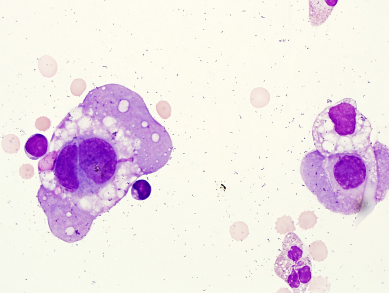
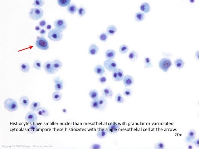
Types of lung cancer and survival rates. Communication with joint wall composition cell lining contents arthrosynovial cyst present continuous mesothelial lining true synovial cells mucinous fluid ganglion cyst may be present discontinuous mesothelial lining flattened pseudosynovial cells mucinous fluid bursitis de novo absent fibrous wall no mesothelial lining fibrinoid necrosis bursa permanent absent continuous. They resemble mesothelial cells but are usually present in smaller numbers figure 1520. The synovial lining cells which make up this membrane produce synovial fluid which lubricates the joints.
The main purpose of these cells is to produce a lubricating fluid that is released between layers providing a slippery non adhesive and protective surface to facilitate intracoelomic movement. The mesothelium is composed of an extensive monolayer of specialized cells mesothelial cells that line the bodys serous cavities and internal organs. By light microscopy synoviocytes are relatively indistinct appearing only as flattened oval dark staining nuclei that usually seem removed from the immediate luminal surface by a fine layer of eosinophilic tissue23 most of the time cytoplasmic borders are not visible.
There are certain cells that line the pleura the thin double layered lining which covers the lungs chest wall and diaphragm which are known as mesothelial cellsother than the pleura mesothelial cells also form a lining around the heart pericardium and the internal surface of the abdomen peritoneum. Mesothelial tissue also surrounds the male. There is a minimal volume of fluid present in a normal joint.
Mesothelial cells in pleural fluid. Identify the morphology of cells found in bronchoalveolar lavage bal. With joint trauma such as infection or inflammation the volume will increase and synovial lining cells may be noted on the cytospin preparation in addition to the cell types.
35035 5 mesothelial cells volume in synovial fluid active part description. Recognize malignant cell morphology in body. Synovial membrane mesothelium lining of the reissners membrane it is now known that the way coronary vessels develop is by the mesenchymal vascular progenitors found within the subepicardium self assembling.
Compare and contrast the morphology of cells found in normal cerebrospinal fluid csf normal pleural fluid normal peritoneal fluid and normal synovial fluid. Lupus erythematosus cells may be present in synovial fluid just as in serous fluid. The pleura thoracal cavity peritoneum abdominal cavity including the mesentery and pericardium heart sac.
Using a direct fluorescent antibody technique bef virus antigen was identified for the first time in synovial pericardial thoracic and abdominal fluids in synovial membranes and epicardium. Distinguish abnormalreactiveinfected cell morphology in fluids. Synovial cells line the synovial cavity and are shed into the cavity.
More From Types Of Lung Cancer And Survival Rates
Incoming Search Terms:
- Http Www Cap Org Apps Docs Committees Hematology Educational Activities 2009 Cmb Pdf Jacob Lee,
- Https Www Sysmex Com Us En Education Webinars Webinar 20handouts W76handout Pdf Jacob Lee,
- Home Jacob Lee,
- Home Jacob Lee,
- Home Jacob Lee,
- Body Fluid Analysis In The Hematology Laboratory Rodak S Hematology Clinical Principles And Applications 5th Ed Jacob Lee,


