Malignant Pleural Effusion Chest X Ray, Elevated Pleural Elastance Common In Malignant Pleural Effusion After Thoracentesis Pulmonology Advisor
Malignant pleural effusion chest x ray Indeed lately has been hunted by consumers around us, maybe one of you personally. Individuals now are accustomed to using the internet in gadgets to view image and video data for inspiration, and according to the name of the article I will discuss about Malignant Pleural Effusion Chest X Ray.
- Management Of Parapneumonic Pleural Effusion In Adults Archivos De Bronconeumologia
- Evaluation Of The Patient With Pleural Effusion Cmaj
- Double Cancer Comprising Malignant Pleural Mesothelioma And Squamous Cell Carcinoma Of The Lung Treated With Radiotherapy A Case Report
- Pleura Chest Wall And Diaphragm Chest Radiology The Essentials 2nd Edition
- Malignant Pleural Effusion Pulmonology Advisor
- Pleural Effusion Undergraduate Diagnostic Imaging Fundamentals
Find, Read, And Discover Malignant Pleural Effusion Chest X Ray, Such Us:
- Overview Of Malignant Pleural Effusion
- Pleural Effusion Chest X Ray Medschool
- Pdf Malignant Pleural Effusion In Breast Cancer 12 Years After Mastectomy That Was Successfully Treated With Endocrine Therapy
- Mmcts
- Https Encrypted Tbn0 Gstatic Com Images Q Tbn 3aand9gctu23kxxwhza8jc Nw0kwcaxx X65he Rcy8lqushp32jgox9q Usqp Cau
- Spider Coloring Pages Halloween
- Emily Walsh Mesothelioma
- Signs Of Pleural Mesothelioma
- Lab Corp Test For Mesothelioma
- Jp Lawn Nagpur
If you are looking for Jp Lawn Nagpur you've come to the ideal place. We have 102 graphics about jp lawn nagpur adding images, photos, pictures, wallpapers, and much more. In these page, we also provide variety of graphics out there. Such as png, jpg, animated gifs, pic art, symbol, blackandwhite, translucent, etc.
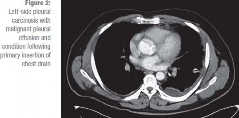
A malignant pleural effusion is often first suspected because of symptoms or findings on a chest x ray or ct scan.

Jp lawn nagpur. However it should be noted that on a routine erect chest x ray as much as 250 600 ml of fluid is required before it becomes evident 6. In the context of a large effusion mediastinal shift toward the side of the effusion should alert the clinician to the possibility of bronchial obstruction which may. It is a fairly common complication in a number of different cancers.
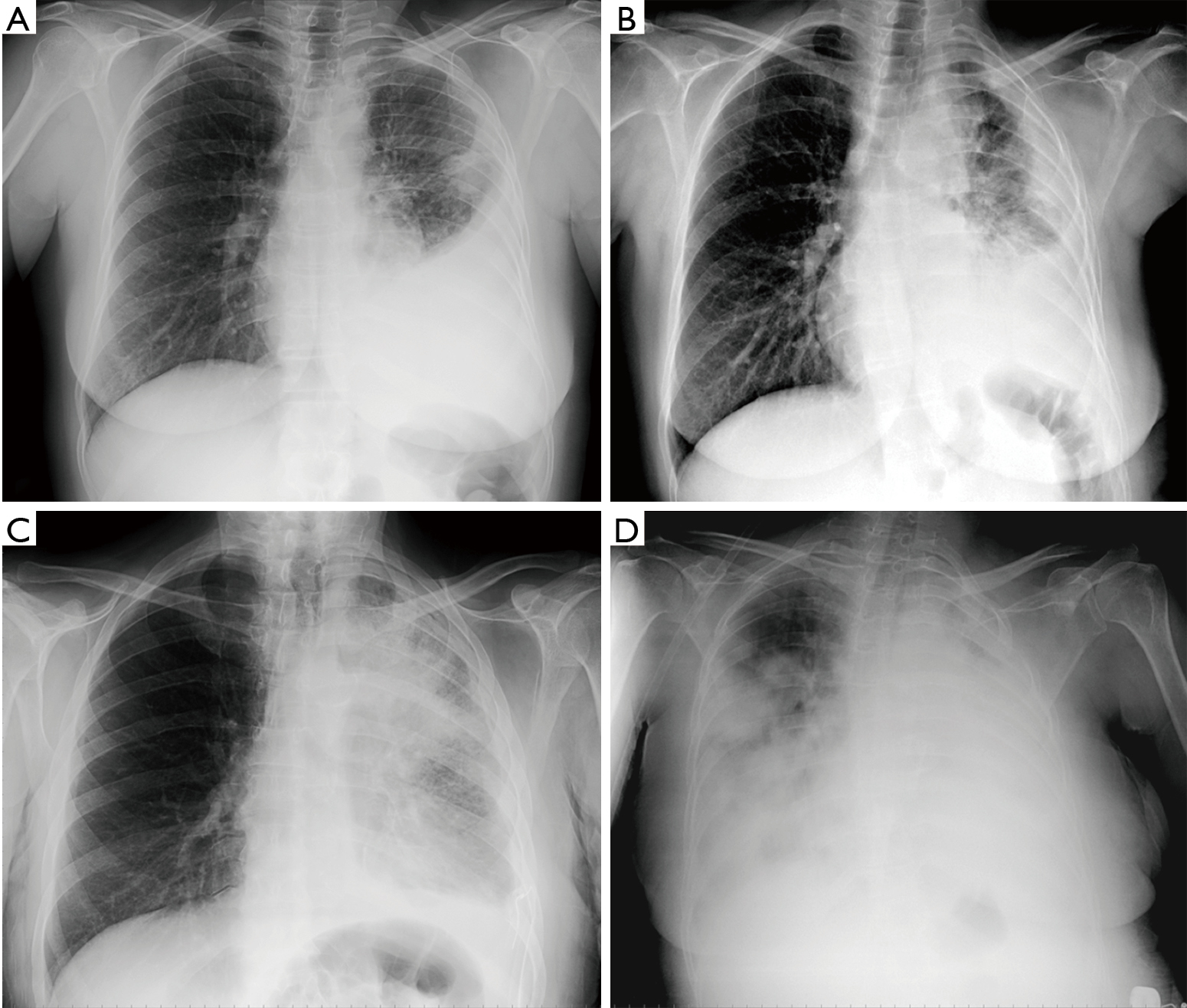
This condition is a sign that the cancer has spread or metastasized to other areas of the body. Chest radiographs are the most commonly used examination to assess for the presence of a pleural effusion. Sixty one patients 587 had massive effusion and 72 patients 692 had mediastinal shift to the opposite side.
Other signs on the chest radiograph may suggest a malignant cause for the effusion. A pleural effusion is a buildup of extra fluid in the space between the lungs and the chest wall. If your doctor suspects a malignant pleural effusion the next step is usually a thoracentesis a procedure in which a needle is inserted through the chest wall into the pleural space to get a sample of the fluid.
A chest x ray can be used to define abnormalities of the lungs such as excessive fluid fluid overload or pulmonary edema fluid around the lung pleural effusion pneumonia bronchitis asthma cysts and cancers. A chest x ray can also detect some abnormalities in the heart aorta and the bones of the thoracic area. A malignant pleural effusion mpe is the build up of fluid and cancer cells that collects between the chest wall and the lung.
The majority of patients who present with a malignant pleural effusion are symptomatic although up to 25 are asymptomatic with an incidental finding of effusion on physical examination or by chest radiography1 dyspnoea is the most common presenting symptom reflecting reduced compliance of the chest wall depression of the ipsilateral. A pleural effusion is usually diagnosed on the basis of medical history and physical exam and confirmed by a chest x rayonce accumulated fluid is more than 300 ml there are usually detectable clinical signs such as decreased movement of the chest on the affected side dullness to percussion over the fluid diminished breath sounds on the affected side decreased vocal resonance and. This area is called the pleural space.
A chest x ray showing a left sided loculated pleural effusion in a patient with mesothelioma. Chest x ray showed right side effusion in 59 patients 567 and 45 had left sided pleural effusion. This can cause you to have chest discomfort as well as feel short of breath.
A lateral decubitus projection is most sensitive able to identify even a small amount of fluid.
More From Jp Lawn Nagpur
- Elsa Coloring Page
- Great Expressions Page Field
- Kelly Lawler Usa Today
- Fluttershy Coloring Pages Equestria Girls
- Doll Coloring Lol Printable Coloring Pages
Incoming Search Terms:
- A Case Of Recurrent Pleural Effusion Can We Think Beyond Tuberculosis And Malignancy Vaishnav B Med J Dy Patil Univ Doll Coloring Lol Printable Coloring Pages,
- Elevated Pleural Elastance Common In Malignant Pleural Effusion After Thoracentesis Pulmonology Advisor Doll Coloring Lol Printable Coloring Pages,
- Pleural Effusion Undergraduate Diagnostic Imaging Fundamentals Doll Coloring Lol Printable Coloring Pages,
- Massive Pleural Effusion Radiology At St Vincent S University Hospital Doll Coloring Lol Printable Coloring Pages,
- Malignant Pleural Effusion Pulmonology Advisor Doll Coloring Lol Printable Coloring Pages,
- Pleural Metastases From Melanoma Radiology Case Radiopaedia Org Doll Coloring Lol Printable Coloring Pages,


