Lymphoma Pleural Effusion Cytology, Effusions Abdominal Thoracic And Pericardial Veterian Key
Lymphoma pleural effusion cytology Indeed lately has been hunted by users around us, perhaps one of you personally. People now are accustomed to using the internet in gadgets to see image and video data for inspiration, and according to the title of this article I will talk about about Lymphoma Pleural Effusion Cytology.
- Liquid Based Cytological Diagnosis Of Achylous Unilateral Bancroftian Pleural Effusion An Uncommon Presentation Of A Common Problem Pandey P Ralli M Agarwal S Agarwal R Med J Dy Patil Vidyapeeth
- Human Herpesvirus 8 Negative Effusion Based Lymphoma With Indolent Clinical Behavior In An Elderly Patient A Case Report And Literature Review
- A Cytology Of Pleural Effusion In January 2014 Showed Large Neoplastic Download Scientific Diagram
- Effusion In Cats Clinician S Brief
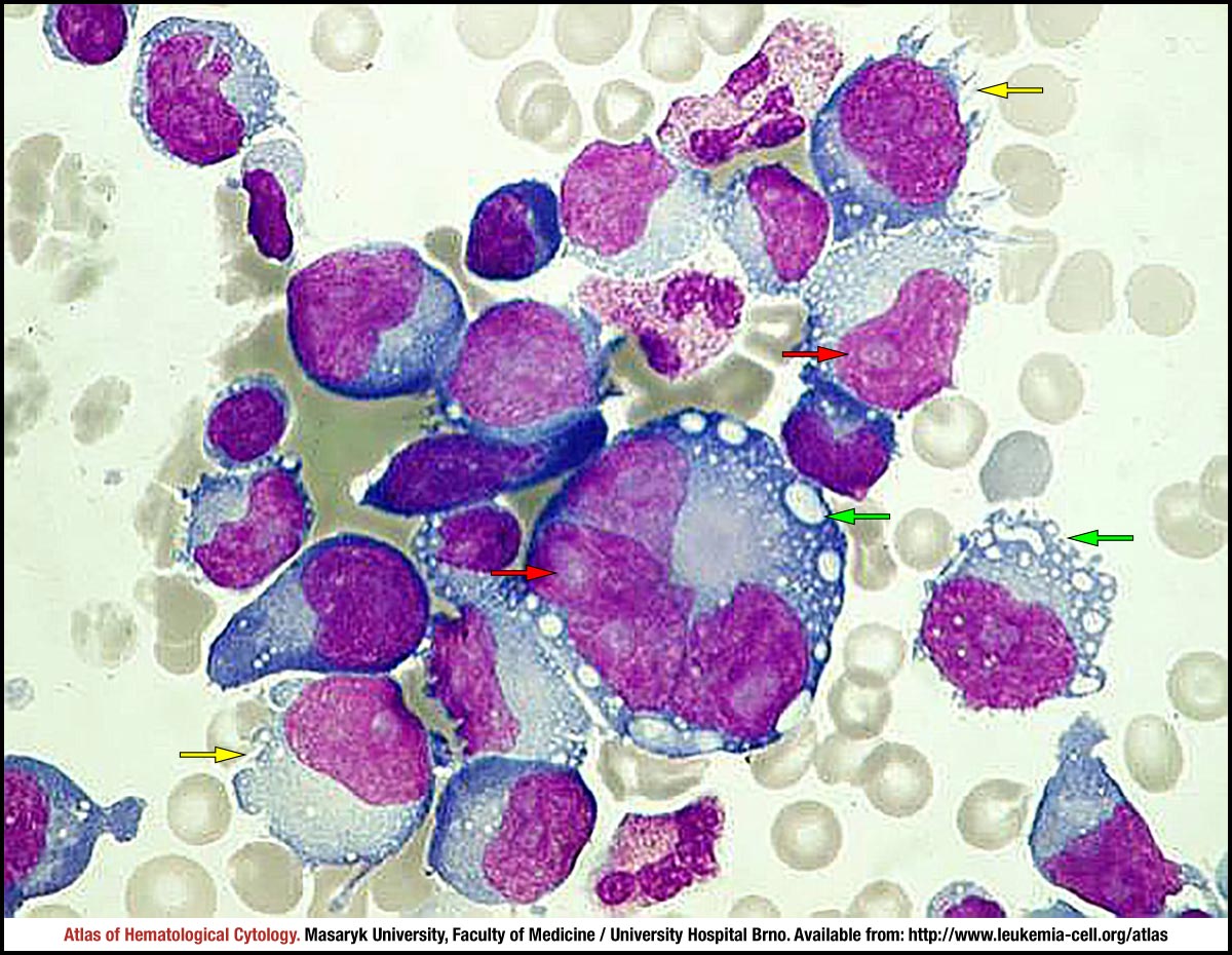
- Peripheral T Cell Lymphoma Not Otherwise Specified Cell Atlas Of Haematological Cytology
- A Comparative Study Of Conventional Cytology And Cell Block Method In The Diagnosis Of Pleural Effusion Assawasaksakul Journal Of Thoracic Disease
Find, Read, And Discover Lymphoma Pleural Effusion Cytology, Such Us:
- Hemorrhagic Pleural Effusion As A First Presentation Of Chronic Lymphocytic Leukemia A Case Report Scialert Responsive Version
- Pleural Myelomatous Involvement In Multiple Myeloma Five Cases Annals Of Saudi Medicine
- Journal Of Pathology And Translational Medicine
- Cytology Of Pleural And Peritoneal Lesions Chapter 5 Practical Pathology Of Serous Membranes
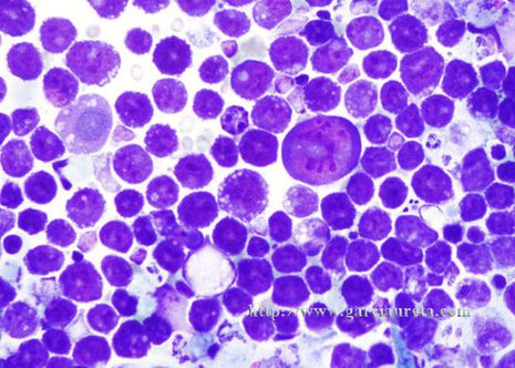
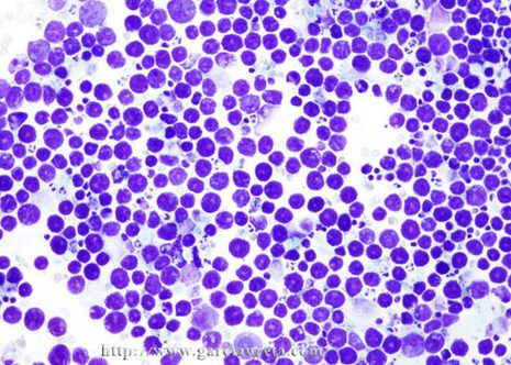
- A And B Pleural Effusion Cytology A Dispersed Lymphoblasts Admixed Download Scientific Diagram
- Key Law Office
- Car Accident Lawyer
- Green Bay Mesothelioma Claim
- Lol Coloring Pages For Girls
- Letter C Coloring Pages
If you re looking for Letter C Coloring Pages you've arrived at the perfect place. We ve got 104 graphics about letter c coloring pages including pictures, photos, pictures, backgrounds, and more. In these page, we additionally provide variety of images available. Such as png, jpg, animated gifs, pic art, symbol, black and white, transparent, etc.
Cytology revealed cells with different morphology and immunophenotype than the histomorphology and immunohistochemistry of the primary infiltrate in the tongue.

Letter c coloring pages. Intrathoracic non hodgkins lymphoma nhl usually presents with roentgenographic evidence of mediastinal lymph node enlargement pulmonary masses pleural effusion and a clinical picture of a systemic disease with lymphadenopathy. Myelomatosis and an encysted pleural effusion myelomacells were found in small numbers among abundant red cells and other nucleated cells inclu ding a few myelocytes and normoblasts. 3 pleural effusion usually occurs as a part of widespread disease chiefly disease associated with mediastinal involvement.
Pleural effusion is a common finding in patients with non hodgkin lymphoma. Pleural effusion from a 79 year old man with a history of lung infiltrates enlarged mediastinal lymph nodes and pleural effusion. The neoplastic cells expressed pax5 in pel and in all b cell lymphomas and cd4 in t cell lymphomas.
The presentation of nhl with pleural effusion as the major roentgenographic abnormality and no clinical peripheral lymphadenopathy or organomegaly is unusual. In hodgkins lymphomas the neoplastic cells expressed cd15 and cd30 markers and the pathognomonic reed stenberg cells were occasionally observed. Acute and chronic leukemias myelodysplastic syndromes are rarely accompanied by pleural involvement.
But the diagnosis could not have been madeon cytological grounds. In the present study nhl was the most common cause of pleural effusion due to lymphoma. Serous effusions are a common complication of lymphomas.
We report a case of pleural involvement with recurrent diffuse large b cell dlbc lymphoma diagnosed by thoracoscopic pleural biopsy. Pleural effusion is not an uncommon finding in patients with nonhodgkins lymphoma nhl with a reported frequency of up to 20. No pleural effusion was observed at that time.
Among lymphoma subtypes t cell neoplasms especially the lymp. Nearly all hematologic malignancies can occasionally present with or develop pleural effusions during the clinical course of disease. 70 year old man with hhv8 ebv pleural effusions j med case rep 2011560 87 year old hiv but hhv8 man with t cell variant arch pathol lab med 20011251246 two cases of hiv hhv8 solid variant without primary effusion lymphoma hum pathol 200233846.
Among the most common disorders are hodgkin and non hodgkin lymphomas with a frequency of 20 to 30 especially if mediastinal involvement is present. Although the frequency of pleural effusion is 20 30 in non hodgkins lymphoma nhl and hodgkins disease hd the involvement of peritoneal and pericardial cavities is uncommon. Fifteen months later a small subcutaneous tumor appeared on the abdomen and pleural fluid was detected.
Thoracoscopy is one of the important diagnostic means especially when there is negative result from fluid cytology. 1 the effusion may be unilateral or bilateral. 1 2 in the majority of these patients the effusion is present at the time of diagnosis.
More From Letter C Coloring Pages
- Heart Colouring Sheet
- Free Lol Surprise Coloring Pages
- Mesothelioma Prevention Tips
- Domingo Garcia Attorney
- Esl Coloring Pages
Incoming Search Terms:
- Cytology Zoom On Twitter Pleural Effusion Of An B Cell Non Hodgkin Lymphoma Cytology Zytologie Cytopathology Esl Coloring Pages,
- Pleural Effusion In A Patient With Lymphoma Malignant Laboratorio De Citologia Laboratorio Esl Coloring Pages,
- Haematology Cytology Atlas Of Organ Biopsies And Exudates Free Medical Atlas Esl Coloring Pages,
- Cytology Of Plasma Cell Rich Effusion In Cases Of Plasma Cell Neoplasm Gochhait D Dey P Verma N J Cytol Esl Coloring Pages,
- Medicina Free Full Text Malignant Pleural Effusion And Its Current Management A Review Html Esl Coloring Pages,
- Peripheral T Cell Lymphoma Not Otherwise Specified Cell Atlas Of Haematological Cytology Esl Coloring Pages,


