Lung Cancer X Ray Of Chest, Chest X Rays Miss Nearly Quarter Of Lung Cancers Review Finds
Lung cancer x ray of chest Indeed recently is being hunted by users around us, perhaps one of you. People are now accustomed to using the net in gadgets to see image and video data for inspiration, and according to the title of the post I will talk about about Lung Cancer X Ray Of Chest.
- Donnelly Centre For Cellular And Biomolecular Research
- Ai Analysis Can Improve Lung Cancer Detection On Chest Radiographs Majalah Kesehatan L Langganan Majalah Kesehatan L Majalah Healthy Times Indonesia
- Chest X Ray For The Diagnosis Of Lung Cancer
- P A Chest X Ray Images Of The Patient With Lung Cancer A Before Download Scientific Diagram
- Chest X Ray And Ct Images Of The Patient With Lung Cancer A Before Download Scientific Diagram
- Cureus Adenocarcinoma Of The Lung Presenting With Intrapulmonary Miliary Metastasis
Find, Read, And Discover Lung Cancer X Ray Of Chest, Such Us:
- Lung Cancer Patients Delayed Treatment Missed Diagnostic Testing Healthmanagement Org
- Lung Cancer Cxr Radiology At St Vincent S University Hospital
- 3
- Yorkshire Cancer Research Announces 7m Investment In Lung Cancer And Early Diagnosis News Yorkshire Cancer Research
- Lung Cancer Thoracic Key
- Jill Lemon Mesothelioma
- Matthew Lawrence Lawrence Brothers
- Is Artex Really Dangerous
- Halloween Pictures For Kids To Color
- Stratified Therapy For Pleural Mesothelioma
If you re searching for Stratified Therapy For Pleural Mesothelioma you've reached the right location. We have 104 images about stratified therapy for pleural mesothelioma including pictures, photos, pictures, wallpapers, and much more. In these page, we additionally provide variety of images available. Such as png, jpg, animated gifs, pic art, symbol, black and white, transparent, etc.

Looking For Lung Cancer With A Yearly X Ray Doesn T Reduce Deaths Shots Health News Npr Stratified Therapy For Pleural Mesothelioma
The consolidation is a result of lunginfarction and bleeding into the alveoli.

Stratified therapy for pleural mesothelioma. Pulmonary embolism resulting in an infarcted area. Hover onoff image to showhide findings. It uses a small amount of radiation to produce an image of your heart lungs and blood vessels.
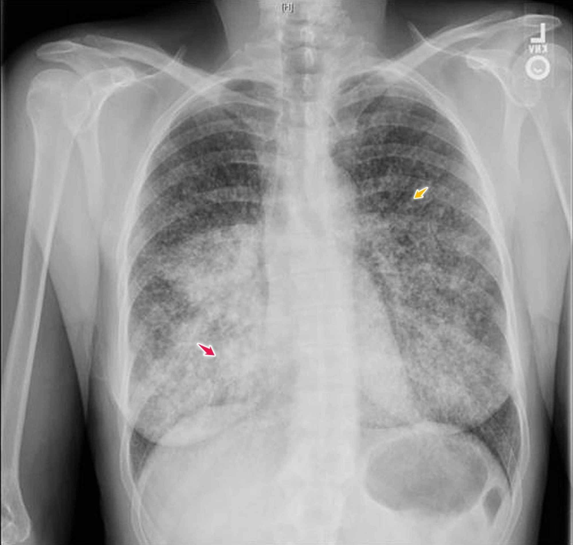
X rays of your chest can be done at imaging centers hospitals or even in some doctors settings. A chest x ray of someone with lung cancer may show a visible mass or nodule. In pulmonar embolism it is not common to see consolidation.
Here a chest x ray of a large cavitating lung cancer which started as a small mass. Lung cancer primary lung cancer or frequently if somewhat incorrectly known as bronchogenic carcinoma is a broad term referring to the main histological subtypes of primary lung malignancies that are mainly linked with inhaled carcinogens with cigarette smoke being a key culprit. Until the lung cancer shows up on a chest x ray the tumor is often too far advanced to be cured.
Lung cancer consolidation. Sometimes your doctor may ask for more tests if the x ray does not clarify completely. Lung cancer consolidation.
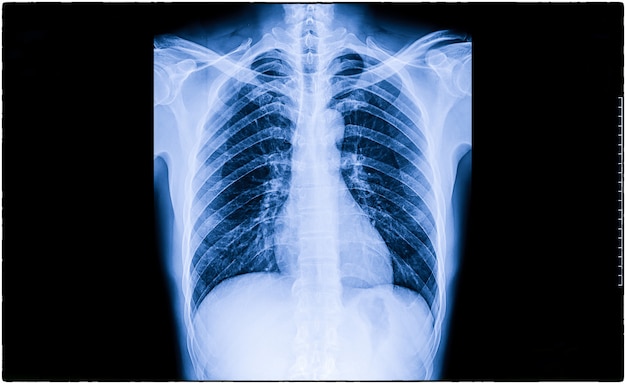
A chest x ray can produce images of your lungs airways heart blood vessels and bones of the chest and spine. Here are the chest x ray appearances of pulmonary metastases from the kidney renal cell carcinoma and gallbladder cholangiocarcinoma. Chest x ray is often the first test your doctor will do if you experience symptoms that match the signs of lung cancer.
It is often the first imaging test your doctor will order if lung or heart disease is suspected. Metastases in the lungs can appear as multiple small or large rounded nodules. This mass will look like a white spot on your lungs while the lung itself will appear black.
Often many things seen on a chest x ray turn out to be treatable problems or artifacts. When diagnosing lung cancer chest x rays do not provide a definitive diagnosis of lung cancers at an early stage when they are more treatable. Pulmonary nodules are small round or oval shaped growth in the.
One of those is a chest x ray. Lung cancerfront x ray image of heart and chest it shows abnormal lung imageology of a femal aged 51 left lung cancer doctor with radiological chest x ray film for medical diagnosis on patients health on asthma lung disease and bone cancer. This article will broadly discuss all the histological subtypes as a group focusing on their common aspects.
If lung cancer is involved. Your doctor uses a chest x ray to.

Chest X Rays Miss Lung Cancer In Almost A Quarter Of Cases Roy Castle Lung Cancer Foundation Stratified Therapy For Pleural Mesothelioma
More From Stratified Therapy For Pleural Mesothelioma
- Show Cause Mesothelioma
- Princess Peach Coloring Sheet
- Halloween Coloring Page For Kids
- Easy Owl Pumpkin Stencil
- Pro Bono Immigration Lawyers Near Me
Incoming Search Terms:
- Normal Chest On X Ray Stock Photo Image Of Medicine 65386600 Pro Bono Immigration Lawyers Near Me,
- Performance Of A Deep Learning Algorithm Compared With Radiologic Interpretation For Lung Cancer Detection On Chest Radiographs In A Health Screening Population Radiology Pro Bono Immigration Lawyers Near Me,
- Diagnostic Imaging Of Lung Cancer European Respiratory Society Pro Bono Immigration Lawyers Near Me,
- Missed Lung Cancer When Where And Why Abstract Europe Pmc Pro Bono Immigration Lawyers Near Me,
- Lung Cancer Pictures X Rays Of Tumors Screening Symptoms And More Pro Bono Immigration Lawyers Near Me,
- Donnelly Centre For Cellular And Biomolecular Research Pro Bono Immigration Lawyers Near Me,


