Lepidic Adenocarcinoma Histology, The Lung Adenocarcinoma Guidelines What To Be Considered By Surgeons Sardenberg Journal Of Thoracic Disease
Lepidic adenocarcinoma histology Indeed lately is being sought by consumers around us, maybe one of you personally. Individuals are now accustomed to using the internet in gadgets to see image and video information for inspiration, and according to the name of the article I will talk about about Lepidic Adenocarcinoma Histology.
- Tumors And Tumor Like Conditions Of The Lung Chapter 18 Silverberg S Principles And Practice Of Surgical Pathology And Cytopathology
- Classic Anatomic Pathology And Lung Cancer Oncohema Key
- Adenocarcinoma Of The Lung From Bac To The Future Insights Into Imaging Full Text
- Pathology Images Gross Pathology Histopathology Histology
- Early Stage Pulmonary Adenocarcinoma T1n0m0 A Clinical Radiological Surgical And Pathological Correlation Of 104 Cases The Md Anderson Cancer Center Experience Modern Pathology
- Carmen Lisievici On Twitter Beautiful Histology Of A Lepidic Adenocarcinoma Of The Lung Stains Are Ck7 And Napsin A Pathology Pulmpath
Find, Read, And Discover Lepidic Adenocarcinoma Histology, Such Us:
- Adenocarcinoma Situ Lung Lepidic Growth Pattern Science Stock Image 1649007451
- Biology Of Invasive Mucinous Adenocarcinoma Of The Lung Cha Translational Lung Cancer Research
- Non Small Cell Lung Cancer Cancer Therapy Advisor
- Aggressive Lung Adenocarcinoma Subtype May Require New Treatment Strategy Memorial Sloan Kettering Cancer Center
- Webpathology Com A Collection Of Surgical Pathology Images
- Vermiculite Vs Asbestos
- Lola Bunny Costume
- Halloween Nail Designs Pictures
- How Do You Diagnose Mesothelioma
- Treat Malignant Pleural Mesothelioma
If you are searching for Treat Malignant Pleural Mesothelioma you've reached the ideal location. We ve got 104 images about treat malignant pleural mesothelioma adding images, photos, pictures, backgrounds, and more. In these page, we additionally provide number of images available. Such as png, jpg, animated gifs, pic art, symbol, blackandwhite, translucent, etc.
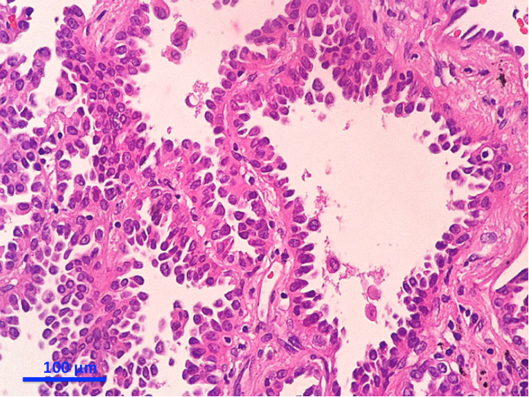
The Lung Adenocarcinoma Guidelines What To Be Considered By Surgeons Sardenberg Journal Of Thoracic Disease Treat Malignant Pleural Mesothelioma
Http Citeseerx Ist Psu Edu Viewdoc Download Doi 10 1 1 663 8305 Rep Rep1 Type Pdf Treat Malignant Pleural Mesothelioma
Or spread through air spaces.
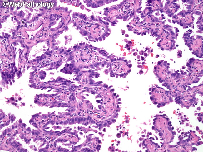
Treat malignant pleural mesothelioma. 3 invasive component greater than 05 cm. Patients with lepidic acinar and mucinous adenocarcinoma had 709 590 and 666 5 year survival respectively and there was no statistically significant difference between them. The subtype is denoted based on the predominant histologic pattern observed.
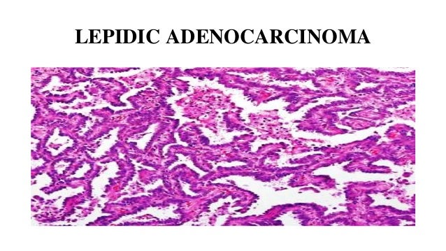
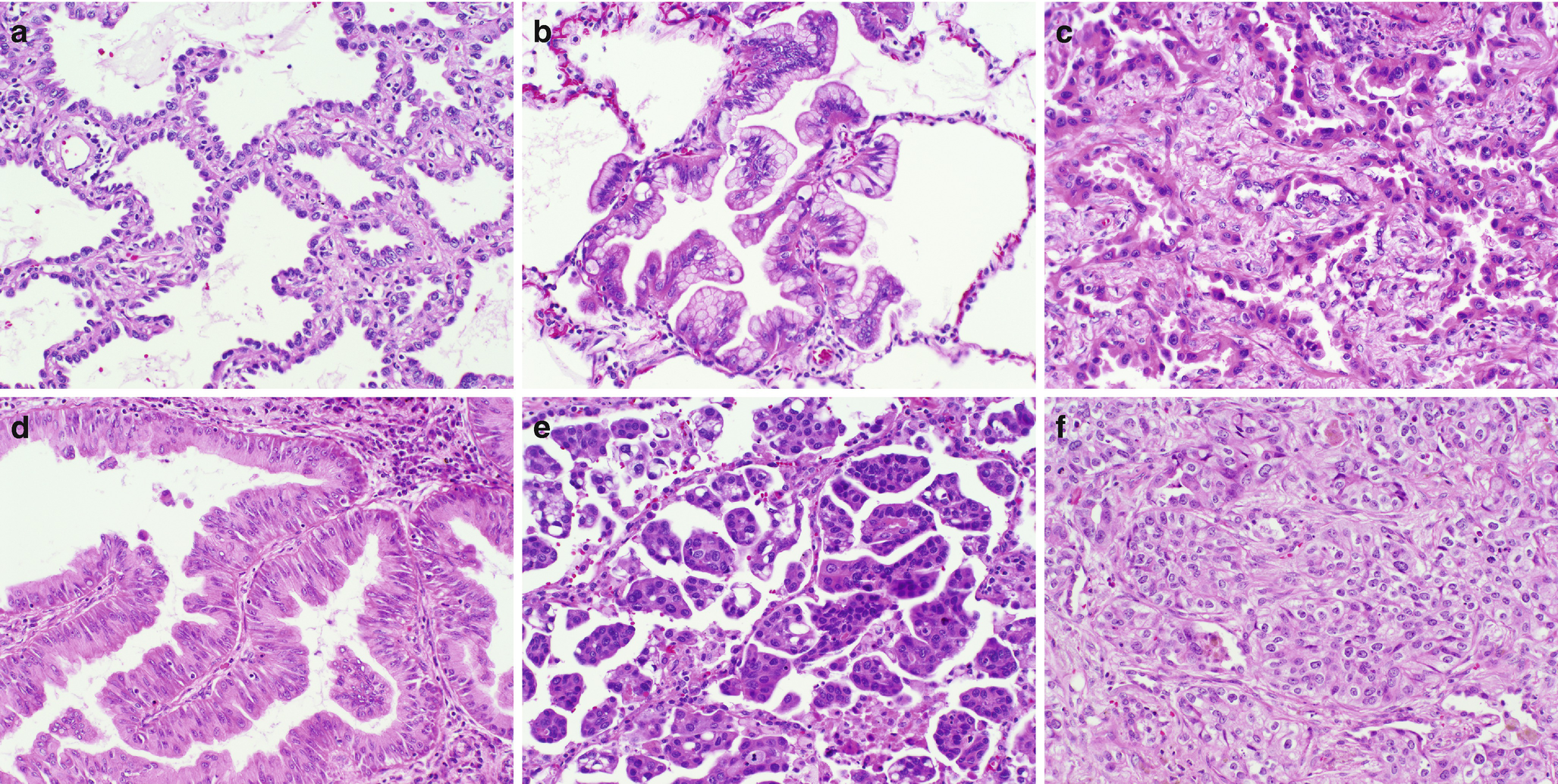
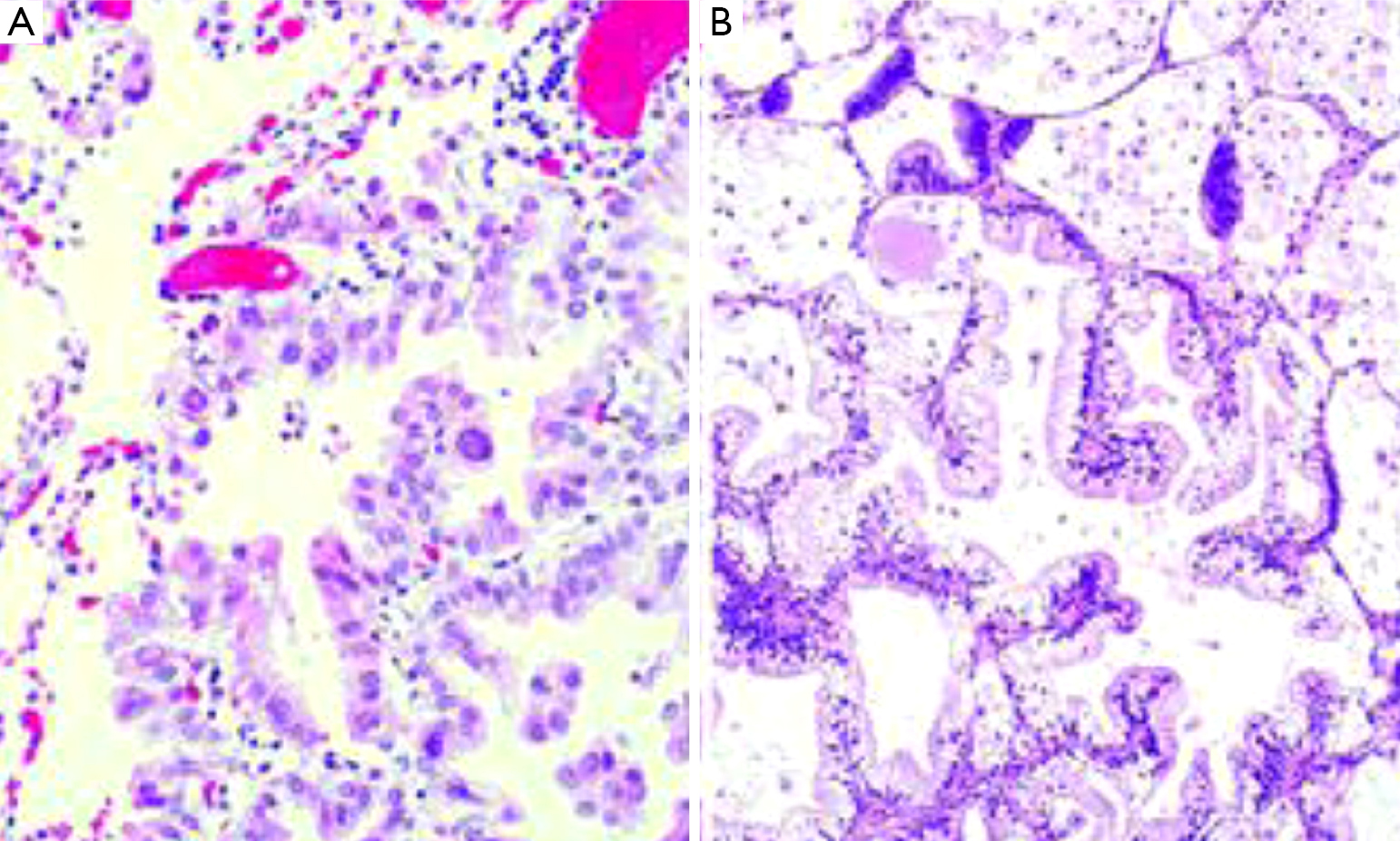
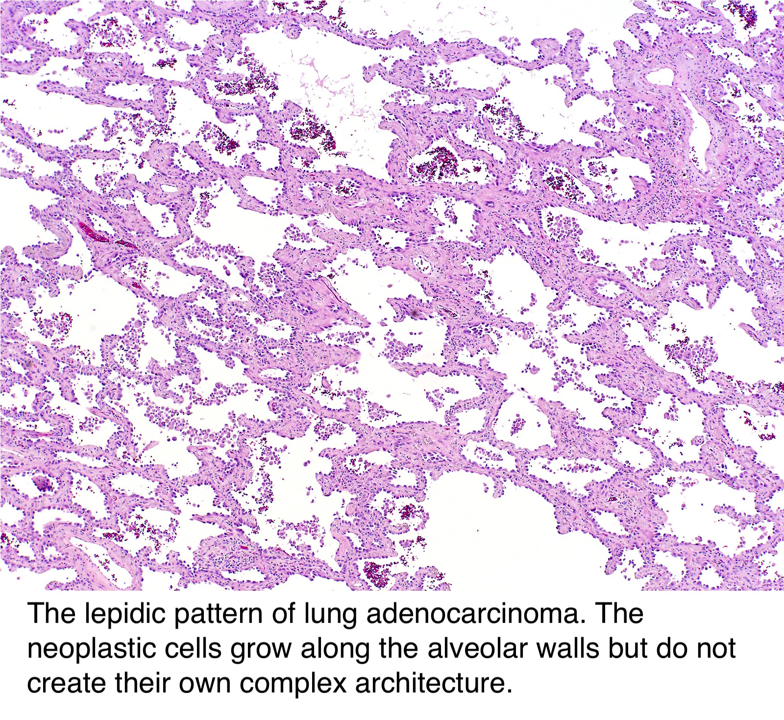
The lepidic growth pattern denotes tumor cells spreading along preexisting alveolar structures although there may be sclerotic thickening of alveolar septa. Adenocarcinoma in situ ais and minimally invasive adenocarcinoma should not be used in the reporting of small biopsies and cytology. Adenocarcinoma in situ minimally invasive adenocarcinoma lepidic predominant adenocarcinoma and invasive mucinous adenocarcinoma are relatively new classification entities which replace the now retired term bronchoalveolar carcinoma bac.
Webpathology is a free educational resource with 10802 high quality pathology images of benign and malignant neoplasms and related entities. Lepidic growth pattern is seen in both lepidic adenocarcinoma as well as in minimally invasive adenocarcinoma mia of lung. Lepidic adenocarcinoma of lung is a histological subtype of pulmonary adenocarcinoma.
Cribriform comedo medullary micropapillary mucinous serrated signet ring cell uncommon subtypes are clear cell adenocarcinoma low grade tubuloglandular adenocarcinoma am j surg pathol 200630. Lepidic growth not as ais. Invasive adenocarcinoma non mucinous.
Glandular neoplasm of the colorectum representing 98 of colonic cancers therefore most details in the general colon carcinoma section pertain to adenocarcinomas. The tumor is diagnosed under a microscope on examination of the cancer cells by a pathologist. The radiographic appearance of these lesions ranges from pure ground glass nodules to large solid masses.
Lung right upper lobe core biopsy. Lepidic adenocarcinoma lepidic growth is commonly seen in lung adenocarcinoma. When it is the predominant growth pattern with additional findings that set it apart from previously.
Whereas patients with solid papillary and micropapillary predominant adenocarcinoma had 410 405 and 00 5 year survival respectively. The diagnosis of lepidic adenocarcinoma is rendered if the tumor shows 1 vascular or pleural invasion.
More From Treat Malignant Pleural Mesothelioma
- Sexy Couples Costume Ideas
- Mesothelioma Asbestos Lawsuit
- Free Printable Trippy Coloring Pages
- Malignant Pleural Mesothelioma Survival Rate
- Printable Detailed Coloring Pages
Incoming Search Terms:
- Histopathological Subtypes And Molecular Alterations In Pulmonary Ade Printable Detailed Coloring Pages,
- Pathology Outlines Adenocarcinoma Overview Printable Detailed Coloring Pages,
- Adenocarcinoma Of The Lung From Bac To The Future Insights Into Imaging Full Text Printable Detailed Coloring Pages,
- Biology Of Invasive Mucinous Adenocarcinoma Of The Lung Cha Translational Lung Cancer Research Printable Detailed Coloring Pages,
- Https Encrypted Tbn0 Gstatic Com Images Q Tbn 3aand9gcquzbquqv Ugexsl5moemhzqviuckv Q Tzgfco7iiqdqvdxtrb Usqp Cau Printable Detailed Coloring Pages,
- Pathology Outlines Adenocarcinoma Overview Printable Detailed Coloring Pages,
