Lepidic Adenocarcinoma Ct, Adenocarcinoma Of The Lung From Bac To The Future Insights Into Imaging Full Text
Lepidic adenocarcinoma ct Indeed recently has been sought by users around us, perhaps one of you. People now are accustomed to using the internet in gadgets to view image and video information for inspiration, and according to the title of the article I will talk about about Lepidic Adenocarcinoma Ct.
- The Iaslc Lung Cancer Staging Project Proposals For Coding T Categories For Subsolid Nodules And Assessment Of Tumor Size In Part Solid Tumors In The Forthcoming Eighth Edition Of The Tnm Classification Of
- Https Encrypted Tbn0 Gstatic Com Images Q Tbn 3aand9gcsqx2etvqnrezzdn807gqxwk3tc0s2ag Fsjuzkhxg Usqp Cau
- Multifocal Adenocarcinoma Of The Lung Multinodular Type Radiology Case Radiopaedia Org
- Ground Glass Opacity Lung Nodules In The Era Of Lung Cancer Ct Screening Radiology Pathology And Clinical Management Cancer Network
- Em Ajr Em Ct Shows Evidence Of Lung Cancer Spread Via Airways
- Slow Growing Lung Cancer As An Emerging Entity From Screening To Clinical Management European Respiratory Society
Find, Read, And Discover Lepidic Adenocarcinoma Ct, Such Us:
- Adenocarcinoma Of The Lung From Bac To The Future Insights Into Imaging Full Text
- Adenocarcinoma Of The Lung From Bac To The Future Insights Into Imaging Full Text
- The Many Faces Of Lung Adenocarcinoma A Pictorial Essay Pascoe 2018 Journal Of Medical Imaging And Radiation Oncology Wiley Online Library
- Former Mucinous Bronchioloalveolar Carcinoma Revisited
- The Many Faces Of Lung Adenocarcinoma A Pictorial Essay Pascoe 2018 Journal Of Medical Imaging And Radiation Oncology Wiley Online Library
- Unicorn Pictures To Colour In
- Haslam Law Firm
- Roblox Colouring Picture
- Get Off My Lawn Sign
- Dr Myers Indiana
If you are searching for Dr Myers Indiana you've come to the right place. We have 104 graphics about dr myers indiana including images, pictures, photos, wallpapers, and more. In these page, we additionally have variety of graphics out there. Such as png, jpg, animated gifs, pic art, logo, blackandwhite, translucent, etc.

Lepidic Predominant Adenocarcinoma Of The Lung Radiology Reference Article Radiopaedia Org Dr Myers Indiana
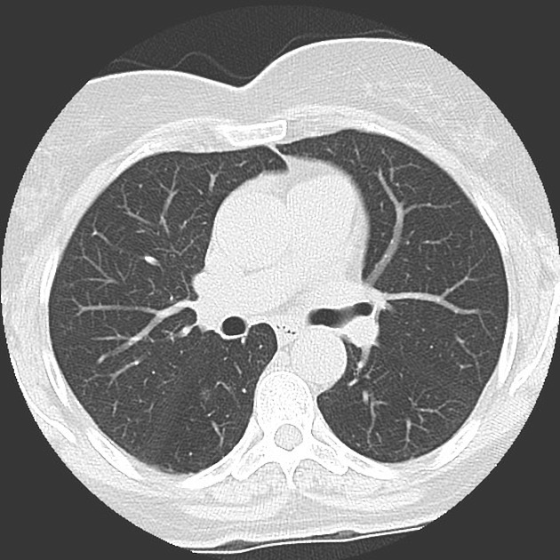
62 year old woman with pure ggn.

Dr myers indiana. Larger diameter of the solid component on ct was also found in lepidic predominant adenocarcinoma compared to minimally invasive adenocarcinoma median 105 vs 2 mm p 005. The tumor is diagnosed under a microscope on examination of the cancer cells by a pathologist. Lepidic predominant adenocarcinoma lpa of the lung formerly known as non mucinous bronchoalveolar carcinoma is a subtype of invasive adenocarcinoma of the lung characterized histologically when the lepidic component comprises the majority of.
Acinar papillary adenocarcinoma had a higher suvmax than lepidic adenocarcinoma with suvmax 14 the optimal cutoff value for differentiation. Lepidic adenocarcinoma of lung is a histological subtype of pulmonary adenocarcinoma. Epub 2017 aug 24.
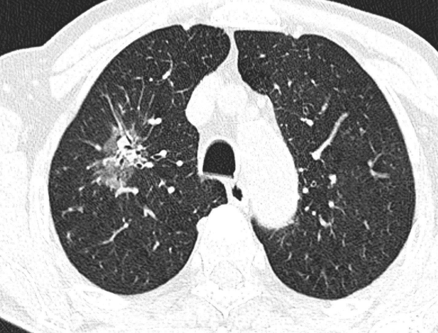
Acinar papillary adenocarcinoma had a higher suv max than lepidic adenocarcinoma median suv max 21 vs 13. Parameter measurements of pure ground glass nodules ggns and part solid ggns on petct fusion and high resolution ct hrct images. The ct appearance is variable but the most typical appearance is a part solid nodule or mass.
Petct plays role in lung adenocarcinoma. The subtype is denoted based on the predominant histologic pattern observed. Lepidic predominant pulmonary lesions lpl.
Lepidic predominant adenocarcinoma of the lung dr daniel j bell and dr yuranga weerakkody et al. An suv max of 14 was the optimal cutoff value for differentiating the growth pattern of adenocarcinoma. For example overtly invasive adenocarcinomas with a lepidic predominant pattern and an invasive component of larger than 5 mm will be classified as lepidic predominant adenocarcinoma.
Ct based distinction from more invasive adenocarcinomas using 3d volumetric density and first order ct texture analysis. Acinar papillary adenocarcinoma had a higher suvmax than lepidic adenocarcinoma with suvmax 14 the optimal cutoff value for differentiation. Adenocarcinoma of the lung is the most common histologic type of lung cancergrouped under the non small cell carcinomas of the lung it is a malignant tumor with glandular differentiation or mucin production expressing in different patterns and degrees of differentiation.
More invasive tumors had higher visual estimated percentage solid component compared to whole lesion measurement on ct p 014. Axial ct image shows a part solid nodule in the right upper lobe arrow.
More From Dr Myers Indiana
- Is All Popcorn Ceiling Asbestos
- Mothra 2019 Coloring Pages
- Funny Scary Pumpkin Carving Patterns
- Pumpkin Carving Ideas Printable
- Train Accident Attorney New York
Incoming Search Terms:
- Adenocarcinoma Of The Lung From Bac To The Future Insights Into Imaging Full Text Train Accident Attorney New York,
- Adenocarcinoma Of The Lung From Bac To The Future Insights Into Imaging Full Text Train Accident Attorney New York,
- The Many Faces Of Lung Adenocarcinoma A Pictorial Essay Pascoe 2018 Journal Of Medical Imaging And Radiation Oncology Wiley Online Library Train Accident Attorney New York,
- The Revised Lung Adenocarcinoma Classification An Imaging Guide Gardiner Journal Of Thoracic Disease Train Accident Attorney New York,
- A Initial Chest Ct Scan A Consolidative Mass In Rml And Rll With Download Scientific Diagram Train Accident Attorney New York,
- Diseases Thoracic Key Train Accident Attorney New York,


