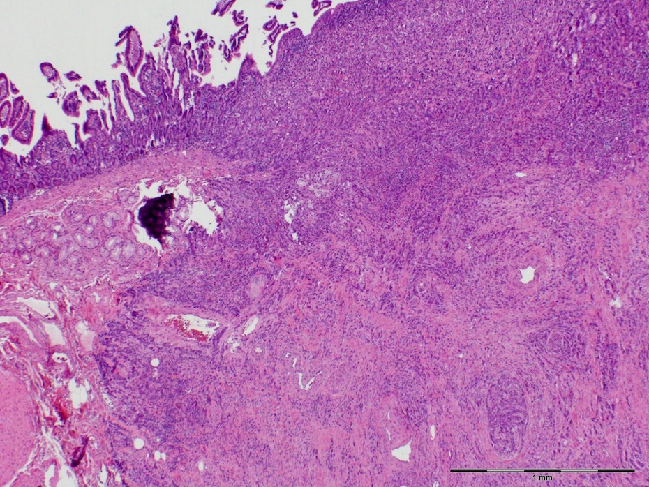
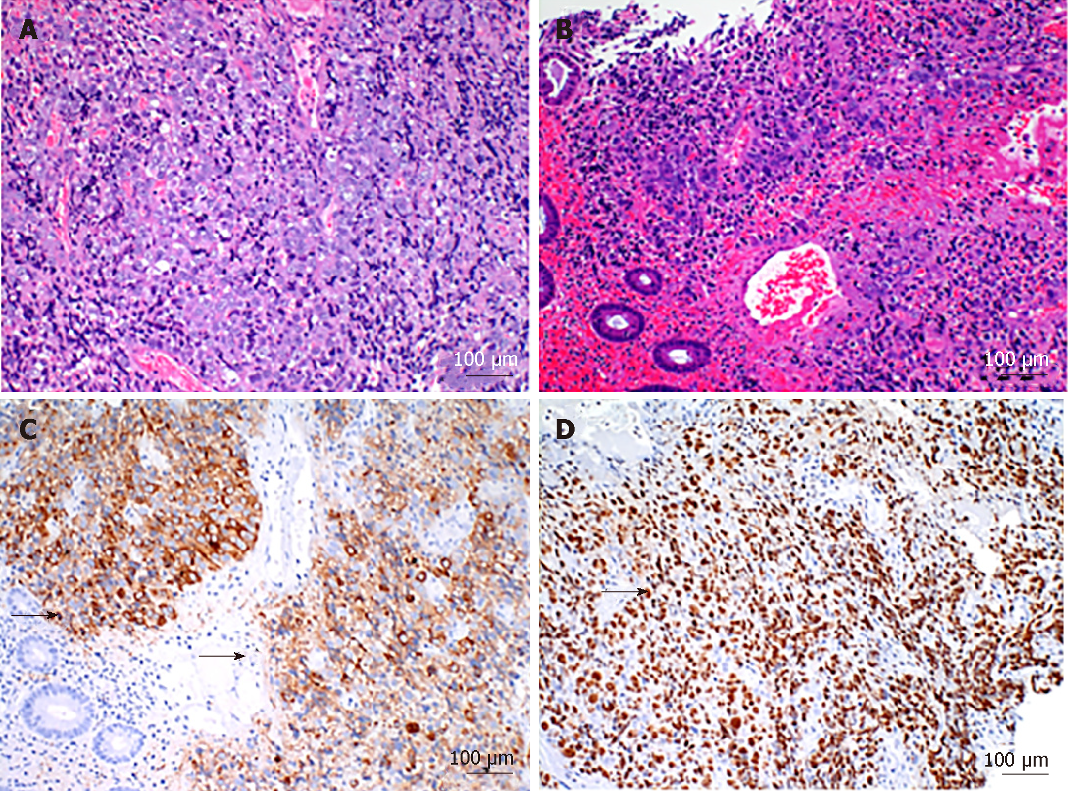
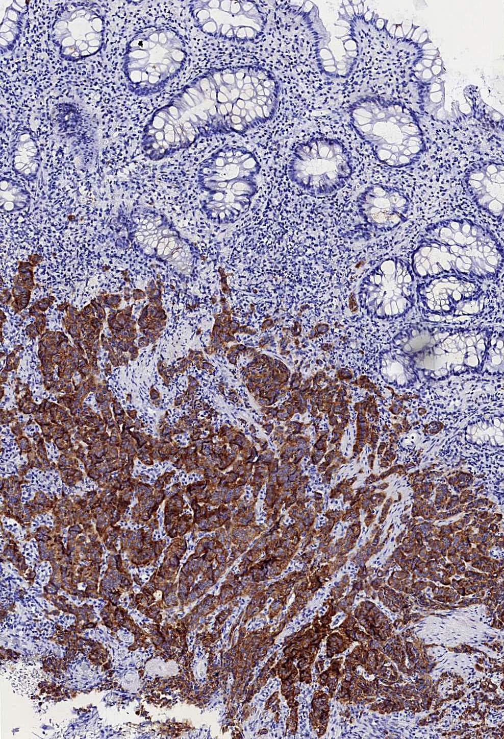
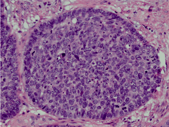
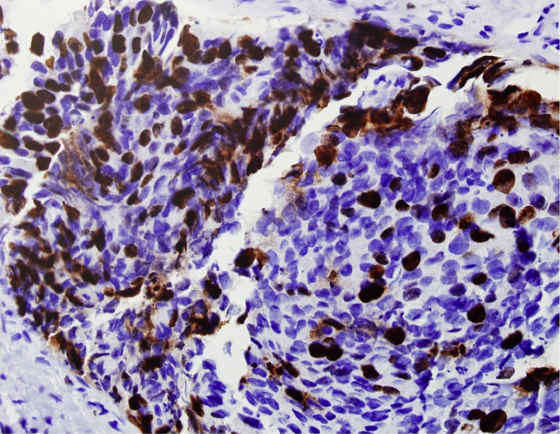
Large Cell Neuroendocrine Carcinoma Colon Pathology Outlines, Https Encrypted Tbn0 Gstatic Com Images Q Tbn 3aand9gcqnqxbrh5zekse9jhi82dh8hwhfcjz5xpv7j1qrinajderrv6wt Usqp Cau
Large cell neuroendocrine carcinoma colon pathology outlines Indeed recently has been hunted by consumers around us, perhaps one of you. Individuals are now accustomed to using the net in gadgets to see video and image data for inspiration, and according to the title of the article I will talk about about Large Cell Neuroendocrine Carcinoma Colon Pathology Outlines.
- Pathology Outlines Neuroendocrine Carcinoma
- Pulmonary Large Cell Neuroendocrine Carcinoma With Adenocarcinoma Like Features Napsin A Expression And Genomic Alterations Modern Pathology
- Pathology Outlines Neuroendocrine Neoplasms General
- Pathology Outlines Neuroendocrine Neoplasms General
- Pathology Outlines Neuroendocrine Neoplasms General
- Pathology Outlines Neuroendocrine Carcinoma
Find, Read, And Discover Large Cell Neuroendocrine Carcinoma Colon Pathology Outlines, Such Us:
- Pathology Outlines Neuroendocrine Neoplasms General
- Colon Tumor Carcinoma Who 2010 Carcinoma Adenocarcinoma
- Pathology Outlines Neuroendocrine Carcinoma
- Pathology Outlines Neuroendocrine Neoplasms General
- Pathology Outlines Neuroendocrine Tumor
- Fritschi Foundry Scholarly Asbestos Mesothelioma
- Primary Peritoneal Mesothelioma Radiology
- Your Lawyers
- Free Printable Fairy Princess Coloring Pages
- Relaxing Coloring Pages Free
If you re looking for Relaxing Coloring Pages Free you've reached the right place. We have 104 graphics about relaxing coloring pages free including images, photos, photographs, wallpapers, and more. In these web page, we also have variety of graphics available. Such as png, jpg, animated gifs, pic art, symbol, blackandwhite, transparent, etc.
Pending neuroendocrine carcinoma.

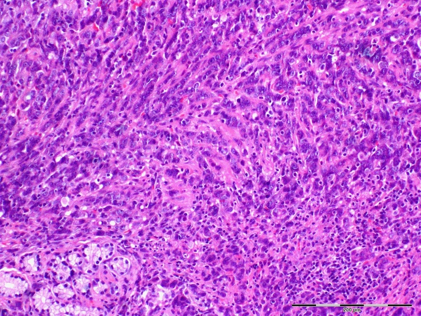
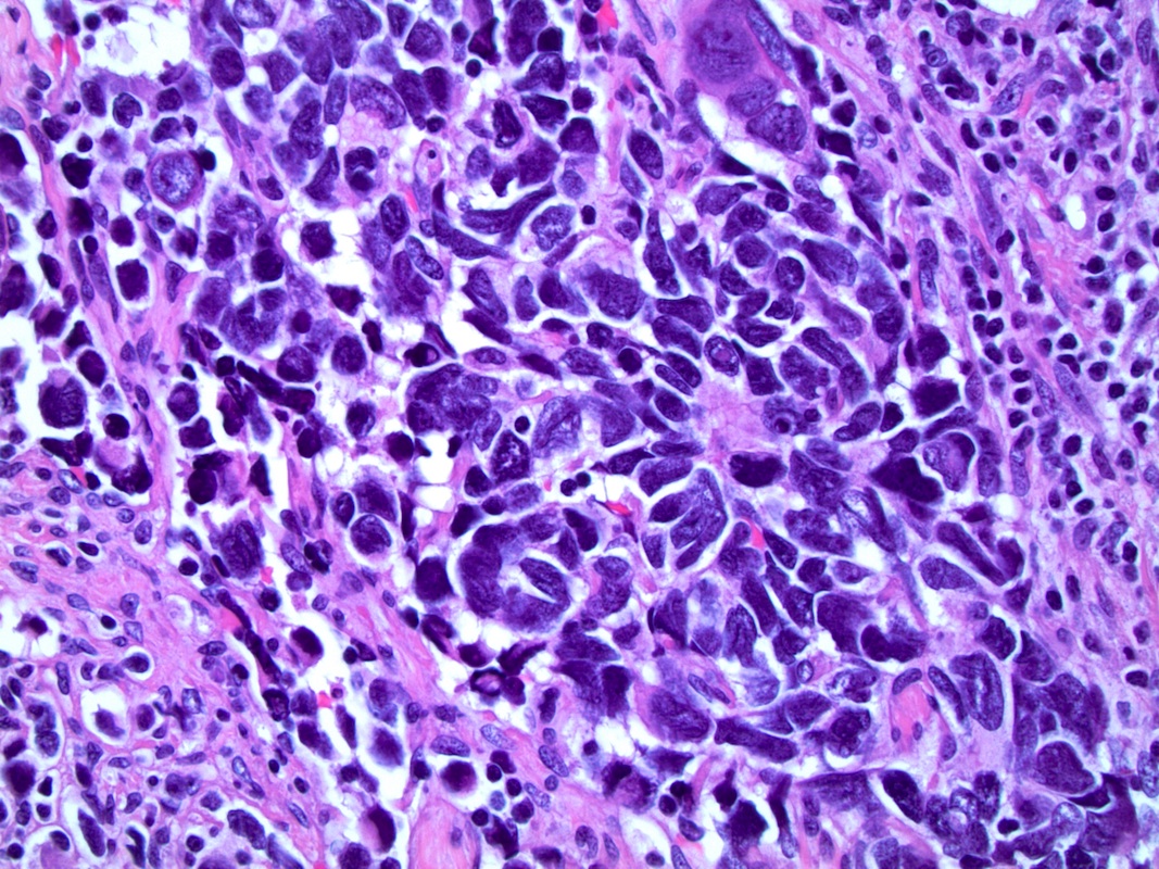
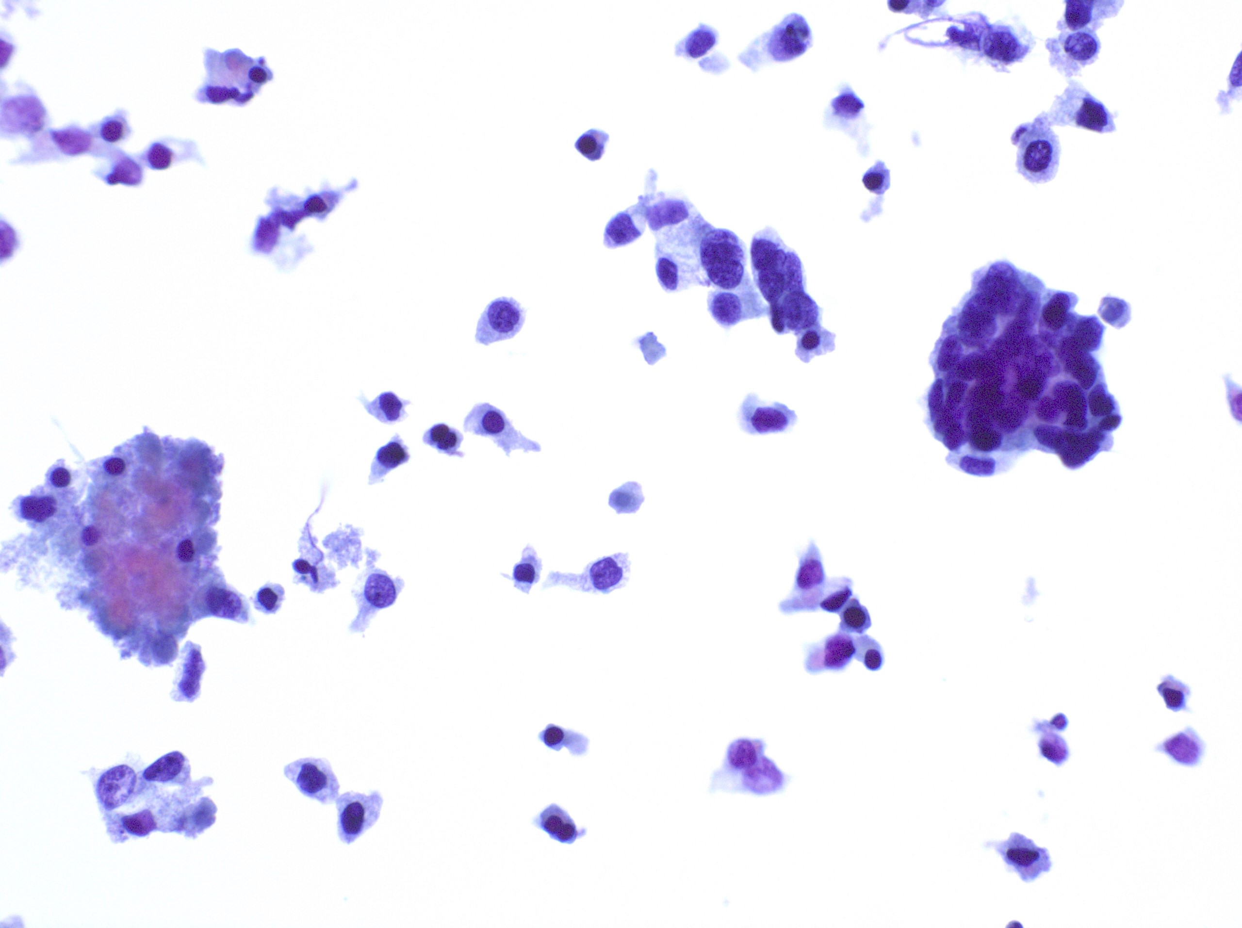
Relaxing coloring pages free. This particular tumor although very likely the same as the neoplasm described first falls short of the. Neuroendocrine carcinomas of the gastrointestinal tract are high grade tumors by definition and typically present at advanced stagethe patients are often younger median age at diagnosis is 55 years than those with colorectal adenocarcinomathe presenting features include abdominal pain obstruction anorexia weight loss rectal bleeding and diarrhea. High grade non small cell carcinoma with neuroendocrine morphology and immunohistochemical markers characterized by 10 mitoses 2mm 2 and extensive necrosis j thorac oncol 2015101243.
Neuroendocrine carcinoma small cell type. Chromogranin and synaptophysin at most focal or scattered. Prognosis for large cell neuroendocrine carcinoma lcnec is poor similar to that of small cell carcinoma.
Colon carcinoma overview posttreatment changes. In the colon neuroendocrine tumors are more frequent in the cecum 696 followed by sigmoid 130 ascending colon 130 and transverse colon 43 arq gastroenterol 200946288 colon proper is the least common site for intestinal wd net. Neuroendocrine carcinoma large cell type.
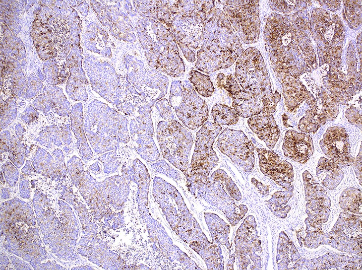
Chromogranin or synaptophysin must be positive in at least 20 50 of cells. This tumor will be classified as large cell carcinoma with neuroendocrine morphology or pattern. A tumor that shows the histopathologic features of a neuroendocrine tumor but which lacks the positive staining for neuroendocrine markers.
Chromatin finely granular stippled. 2 mitoses2 mm2 or 3 ki 67 and 20 mitoses2 mm2 or 20 ki 67. Poorly differentiated neuroendocrine carcinoma large cell type.
17 colon 14 rectum 6 anal canal and 1 appendix. The tumor demonstrates large cell neuroendocrine carcinoma morphology. Poorly differentiated neuroendocrine carcinoma see synoptic report and note.
Tumors were located as follows. Serrated adenoma shigella signet ring cell solitary fibrous tumor solitary rectal ulcer syndrome spirochetosis squamous cell staging carcinoma staging neuroendocrine stercoral ulcer strongyloides stercoralis syphilis tactile corpuscle. Pathology was reviewed and tumors were categorized as small cell carcinoma n 22 or large cell neuroendocrine carcinoma n.
The diagnosis of neuroendocrine carcinoma was suggested preoperatively from tissue biopsy in 593 percent 1627 of patients evaluable.
More From Relaxing Coloring Pages Free
- Easy Pictures To Colour In
- Lamborghini Coloring Pages For Boys
- Halloween Day 2018
- Meyer Park Fort Smith Ar
- Mesothelioma Government Compensation Uk
Incoming Search Terms:
- Https Encrypted Tbn0 Gstatic Com Images Q Tbn 3aand9gcqzbn Svmpfuxbvn0kwk13qrnw3kkg4mkfrnfl52ip2aogekbe Usqp Cau Mesothelioma Government Compensation Uk,
- Pathology Outlines Neuroendocrine Neoplasms General Mesothelioma Government Compensation Uk,
- Pathology Outlines Neuroendocrine Neoplasms General Mesothelioma Government Compensation Uk,
- Pathology Outlines Large Cell Mesothelioma Government Compensation Uk,
- Pathology Outlines Neuroendocrine Tumor Mesothelioma Government Compensation Uk,
- Pathology Outlines Neuroendocrine Tumor Mesothelioma Government Compensation Uk,
