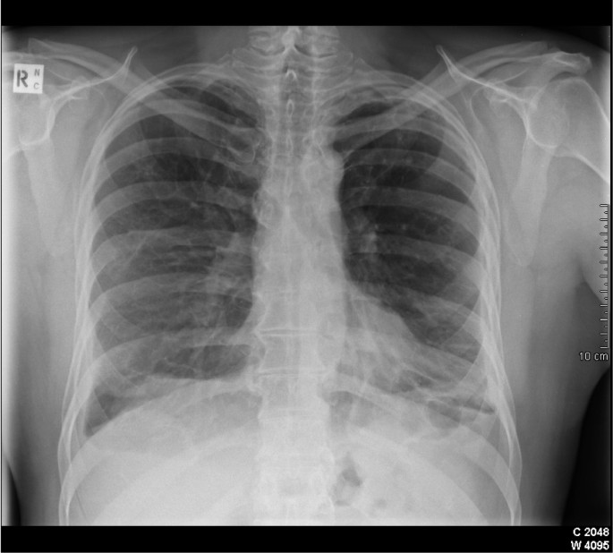
Focal Pleural Thickening Radiology, Https Encrypted Tbn0 Gstatic Com Images Q Tbn 3aand9gcrhefpkbabcdkcnpr06l5pzxaisl4ojyreu5ycqmwkaujl3wgmo Usqp Cau
Focal pleural thickening radiology Indeed lately is being sought by users around us, maybe one of you. People now are accustomed to using the net in gadgets to see image and video data for inspiration, and according to the title of this post I will discuss about Focal Pleural Thickening Radiology.
- Https Www Birpublications Org Doi Pdf 10 1259 Bjr 20150792
- Radiological Review Of Pleural Tumors Sureka B Thukral Bb Mittal Mk Mittal A Sinha M Indian J Radiol Imaging
- Https Www Ajronline Org Doi Pdfplus 10 2214 Ajr 154 3 2106209
- Incidental And Underreported Pleural Plaques At Chest Ct Do Not Miss Them Asbestos Exposure Still Exists
- Imaging Of The Pleura Helm 2010 Journal Of Magnetic Resonance Imaging Wiley Online Library
- A Systematic Approach To Chest Radiographic Analysis Springerlink
Find, Read, And Discover Focal Pleural Thickening Radiology, Such Us:
- Tumorlike Conditions Of The Pleura Radiographics
- Imaging Characteristics Of Pleural Tumours Springerlink
- Epos Trade
- 02 Pleural Disease 2019 Radiology
- Benign Pleural Thickening Radiology Key
- Toy Story Printable Pictures
- Mesothelioma Brain Tumor
- What Is Symptoms Of Mesothelioma
- Japanese Anime Coloring Book
- Space Coloring Pictures
If you re searching for Space Coloring Pictures you've arrived at the right location. We ve got 104 images about space coloring pictures adding images, pictures, photos, backgrounds, and more. In these web page, we also have variety of images out there. Such as png, jpg, animated gifs, pic art, symbol, blackandwhite, transparent, etc.
According to etiology it may be classified as.

Space coloring pictures. 1 however the diagnosis of pt may be problematic and medico legal issues may occur. Diffuse tumorlike conditions of the pleura include diffuse pleural thickening and rare conditions such as erdheim chester disease and diffuse pulmonary. Abstract pleural thickening has a variety of causes and often must be distinguished from pleural masses while pleural calcifications are frequently the result of chronic infections including bacterial or tuberculous empyema.
Calcification or other high attenuation within the pleural space is commonly due to asbestos exposure chemical pleurodesis and remote trauma or infection. Extrapleural hematoma is a nonpleural mimic of pleural tumor and shares some imaging features with focal tumorlike conditions of the pleura despite residing in the extrapleural space. Pleural fibrosis has a number of causes and is the outcome of many pleural diseases and a potential complication of every inflammatory condition that affects the lungs.
The authors explore the options learning points a 77 year old man presented with left sided chest and back pain that did not respond to simple analgesics. That is located within the interlobular septa and along the pleural linings. Radiology department of the rijnland hospital leiderdorp and the academical medical centre amsterdam the netherlands.
As pleural thickening can have a benign or malignant cause use of the appropriate imaging techniques is crucial to a correct diagnosis. Benign pleural thickening caused by fibrosis is the second most common pleural abnormality the most common one being effusion. In 50 of patients the septal thickening is focal or unilateral.
Pleural thickening may be focal or diffuse benign or malignant with characteristic imaging features that can narrow the differential diagnosis table 1. The pleural plaques of asbestos may be localized soft tissue but frequently calcify with a characteristic radiologic appearance on both chest x ray and ct. The analysis of the location and shape of the pt as well as.
To propose an imaging strategy and propose an explanation for their mechanism of formation. 1 however the diagnosis of pt may be problematic and medico legal issues may occur. It can occur with both benign and malignant pleural disease.
Numerous causes of focal pleural thickening pt may be seen in routine chest ct. The pleura show a variety of patterns of fibrosis. Diagnosis of pleural plaques pps is usually feasible especially when a typical appearance is associated with a history of previous asbestos exposure.
He had a history of atrial fibrillation and was taking warfarin. To describe the characteristics of reversible focal pleural thickenings pts mimicking real plaques that firstly suggest asbestos exposure or pleural metastasis. Numerous causes of focal pleural thickening pt may be seen in routine chest ct.
The analysis of the location and shape of the pt as well as associated.
More From Space Coloring Pictures
- Kel Law Firm
- Mesothelioma Treatment Mesothelioma Treatment Mayo Clinic
- Michael Jordan Coloring Pages
- Mesothelioma Malayalam Meaning
- Creative Law Firm Names
Incoming Search Terms:
- Https Www Birpublications Org Doi Pdf 10 1259 Bjr 20150792 Creative Law Firm Names,
- Pleural Fibrosis And Calcification Pulmonary Disorders Msd Manual Professional Edition Creative Law Firm Names,
- Is Transthoracic Ultrasound Tus A Reliable Predictor Of The Nature Of Pleural And Peripheral Pulmonary Lesions Correlation With Cyto Histological Findings Egyptian Journal Of Radiology And Nuclear Medicine Full Text Creative Law Firm Names,
- Pleura Chest Wall And Diaphragm Thoracic Key Creative Law Firm Names,
- Pdf Clinical Consequences Of Asbestos Related Diffuse Pleural Thickening A Review Creative Law Firm Names,
- Pleural Thickening Radiology Reference Article Radiopaedia Org Creative Law Firm Names,


