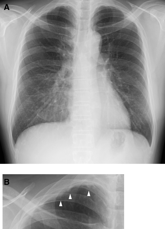
Focal Pleural Thickening Ct, Pleura Chest Wall And Diaphragm Chest Radiology The Essentials 2nd Edition
Focal pleural thickening ct Indeed lately is being hunted by users around us, perhaps one of you personally. People now are accustomed to using the internet in gadgets to view video and image information for inspiration, and according to the name of this post I will talk about about Focal Pleural Thickening Ct.
- Benign Pleural Thickening Radiology Key
- Https Encrypted Tbn0 Gstatic Com Images Q Tbn 3aand9gcqm2wnxdnjwxjiarbr9ietjmckxbf5eolhcrkbcawdyim9zjp0n Usqp Cau
- Radiological Imaging Of Pleural Diseases
- Pleural Thickening Radiology Reference Article Radiopaedia Org
- 2
- Focal Dependent Pleural Thickening At Mdct Pleural Lesion Or Functional Abnormality Sciencedirect
Find, Read, And Discover Focal Pleural Thickening Ct, Such Us:
- Investigating Pleural Thickening The Bmj
- Radiological Imaging Of Pleural Diseases
- Comparative Interpretation Of Ct And Standard Radiography Of The Pleura
- Pleura Chest Wall And Diaphragm Radiology Key
- Focal Pleural Tumorlike Conditions Nodules And Masses Beyond Mesotheliomas And Metastasis Sciencedirect
- Disney Store Costumes
- Education Law Clipart
- Latest News On Mesothelioma
- Nurse Pumpkin Carving Patterns
- Malignant Mesothelioma Histopathology Features
If you are looking for Malignant Mesothelioma Histopathology Features you've come to the ideal location. We ve got 104 graphics about malignant mesothelioma histopathology features adding images, photos, pictures, wallpapers, and much more. In such page, we additionally have variety of images available. Such as png, jpg, animated gifs, pic art, symbol, blackandwhite, translucent, etc.
Https Www Ajronline Org Doi Pdfplus 10 2214 Ajr 154 3 2106209 Malignant Mesothelioma Histopathology Features

Radiological Review Of Pleural Tumors Sureka B Thukral Bb Mittal Mk Mittal A Sinha M Indian J Radiol Imaging Malignant Mesothelioma Histopathology Features
The apex of the lung was the most frequently affected area additional file 1.

Malignant mesothelioma histopathology features. 2amore than half of the cases were bilateral and 357 involved thickening on the. The presence of a thin layer of normal lung density intervening between some of these subpleural opacities and the focal pts confirms the parenchymal nature of the former opacities. Numerous causes of focal pleural thickening pt may be seen in routine chest ct.
According to etiology it may be classified as. Focal pleural thickening mimicking pleural plaques on chest computed tomography. It can occur with both benign and malignant pleural disease.
The analysis of the location and shape of the pt as well as associated. However this feature may also be presentalbeit less commonlywith malignancy 18 19. Plaques typically have edges that are thicker than the central portion fig.
Pleural thickening was found predominantly at the apex of the right lung. 1 however the diagnosis of pt may be problematic and medico legal issues may occur. On ct a pleural plaque is defined as a discontinuous soft tissue focal thickening of the pleural surface with or without foci of calcification.
It is a component of the loose connective tissue of the endothoracic fascia and is most abundant along the posterolateral aspects of the 4 th through to 8 th ribs bilaterally. It can occur in various locations but typically occurs along the chest wall. Table s2pleural thickening involving the apical area of either lung was defined as an apical cap which accounted for 922 n 836907 of the cases fig.
The thick soft tissue density at the chest wall lung interface on the axial ct images sometimes do not truly suggest pleural thickening on ct. 1 however the diagnosis of pt may be problematic and medico legal issues may occur. The analysis of the location and shape of the pt as well as.
Pleural thickening is a descriptive term given to describe any form of thickening involving either the parietal or visceral pleura. Thickening of the extrapleural fat as detected on ct images is indicative of a chronic benign cause of the adjacent pleural thickening. The hypothesis of the pleural nature of the underlying thickening is supported by the large base of implantation of the focal thickening with the pleura.
Numerous causes of focal pleural thickening pt may be seen in routine chest ct. Physiological pleural fluid accumulation or dependent. Diagnosis of pleural plaques pps is usually feasible especially when a typical appearance is associated with a history of previous asbestos exposure.

Imaging Of The Pleura Helm 2010 Journal Of Magnetic Resonance Imaging Wiley Online Library Malignant Mesothelioma Histopathology Features
More From Malignant Mesothelioma Histopathology Features
- Cat Coloring Pages For Adults
- Dinosaur Carnotaurus Coloring Pages
- Cute Snake Coloring Pages
- January Calendar Coloring Pages
- Martin Om28jm
Incoming Search Terms:
- 2 Martin Om28jm,
- Diagnostic Imaging And Workup Of Malignant Pleural Mesothelioma Martin Om28jm,
- Pleural Thickening Radiology Reference Article Radiopaedia Org Martin Om28jm,
- Https Www Ajronline Org Doi Pdfplus 10 2214 Ajr 154 3 2106209 Martin Om28jm,
- Radiological Review Of Pleural Tumors Sureka B Thukral Bb Mittal Mk Mittal A Sinha M Indian J Radiol Imaging Martin Om28jm,
- Benign Pleural Thickening Radiology Key Martin Om28jm,


