Chrysotile Asbestos Under Microscope, Chrysotile Fibers Of Asbestos Under Plm Microscope Tiny Stories Asbestos Science Nature
Chrysotile asbestos under microscope Indeed lately has been hunted by consumers around us, perhaps one of you personally. Individuals now are accustomed to using the internet in gadgets to view image and video information for inspiration, and according to the name of the article I will talk about about Chrysotile Asbestos Under Microscope.
- What Does Asbestos Look Like How To Recognise Asbestos Materials
- A B Asbestos Bodies Observed Under The Optical Microscope C Download Scientific Diagram
- A Fibers And Large Fiber With Splayed End Of Raw Chrysotile Imaged In Download Scientific Diagram
- Sem Chrysotile
- What Is Asbestos National Asbestos Helpline National Asbestos Helpline
- Study On The Thermal Decomposition Of Chrysotile Asbestos Springerlink
Find, Read, And Discover Chrysotile Asbestos Under Microscope, Such Us:
- Asbestos Testing As4964 Asbestos Testing
- Exposure To Asbestos And Exposure Monitoring At Helsinki Asbestos 2014
- Details Public Health Image Library Phil Health Images Image Scanning Electron Microscope
- Fatal Asbestos Exposure Level
- Asbestos Type Has No Effect On Mesothelioma Latency Period
- Mesothelioma Treatment Alternative Treatment
- Peppa Pig Printables Free
- Noncalcified Pleural Plaques
- Free Christmas Coloring Pages For Adults
- Dora Coloring Pages Free Printable
If you re searching for Dora Coloring Pages Free Printable you've arrived at the perfect place. We have 100 graphics about dora coloring pages free printable including images, photos, pictures, wallpapers, and more. In these webpage, we additionally have number of graphics available. Such as png, jpg, animated gifs, pic art, symbol, blackandwhite, transparent, etc.
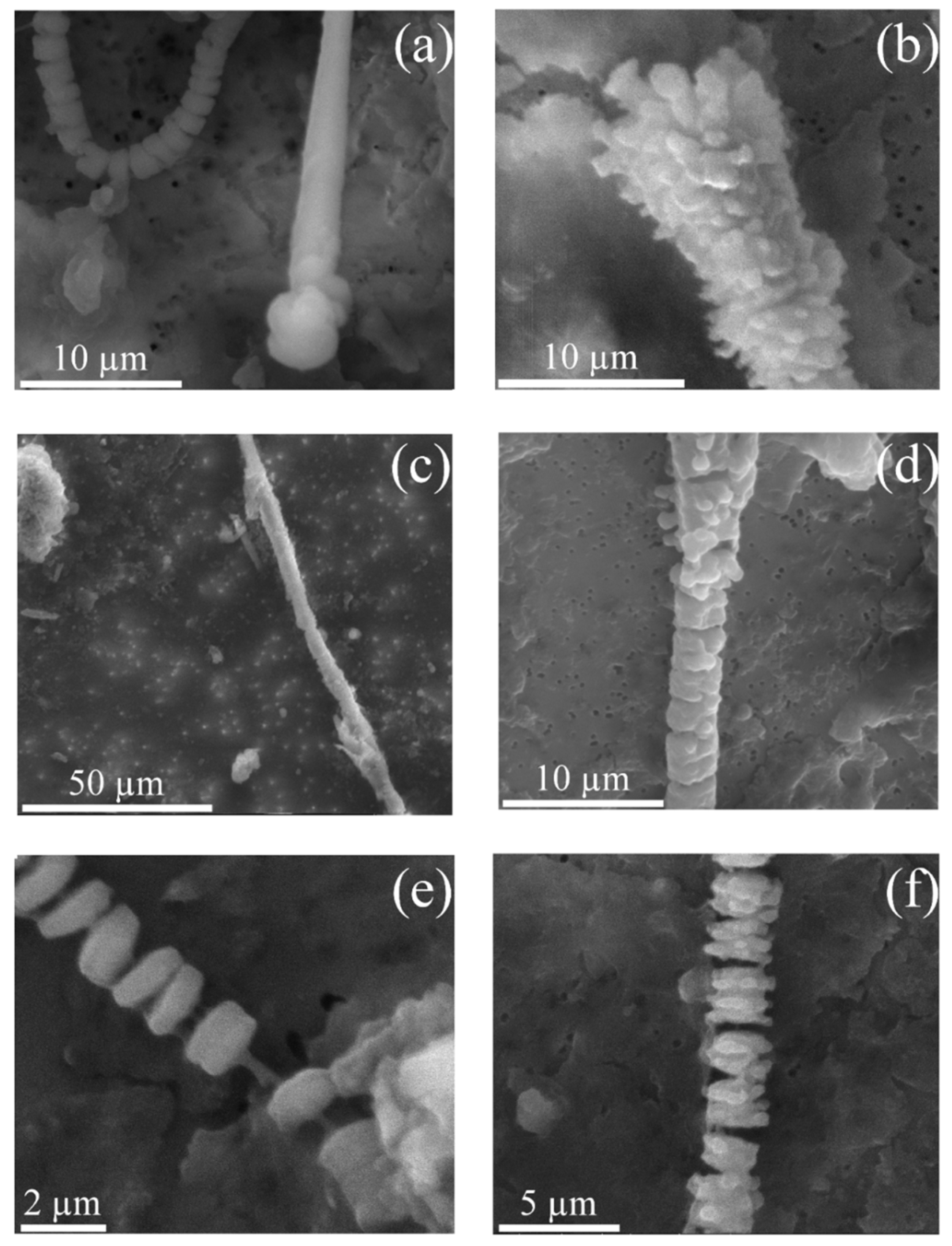
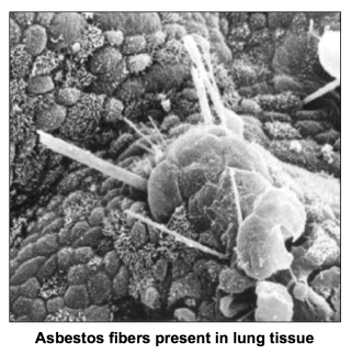
Electron microscope characteristics of inhaled chrysotile asbestos fibre.

Dora coloring pages free printable. Specimens from 300 lungs have been examined under the electron microscope to determine the morphologyanddiffraction characteristics ofany chrysotile asbestos they contained. This structure made it particularly useful in the manufacture of fire resistant clothing and textiles in conveyor belts exposed to high heat environments and in construction materials requiring strength. A low powered stereo microscope eg 8 to 40 magnification is required for the initial search for fibres.
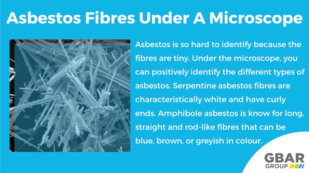
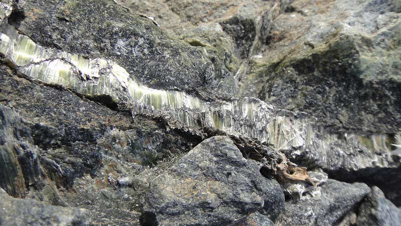
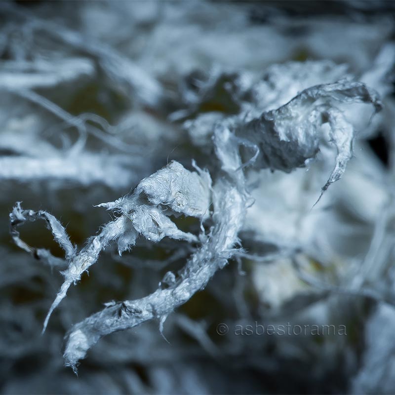
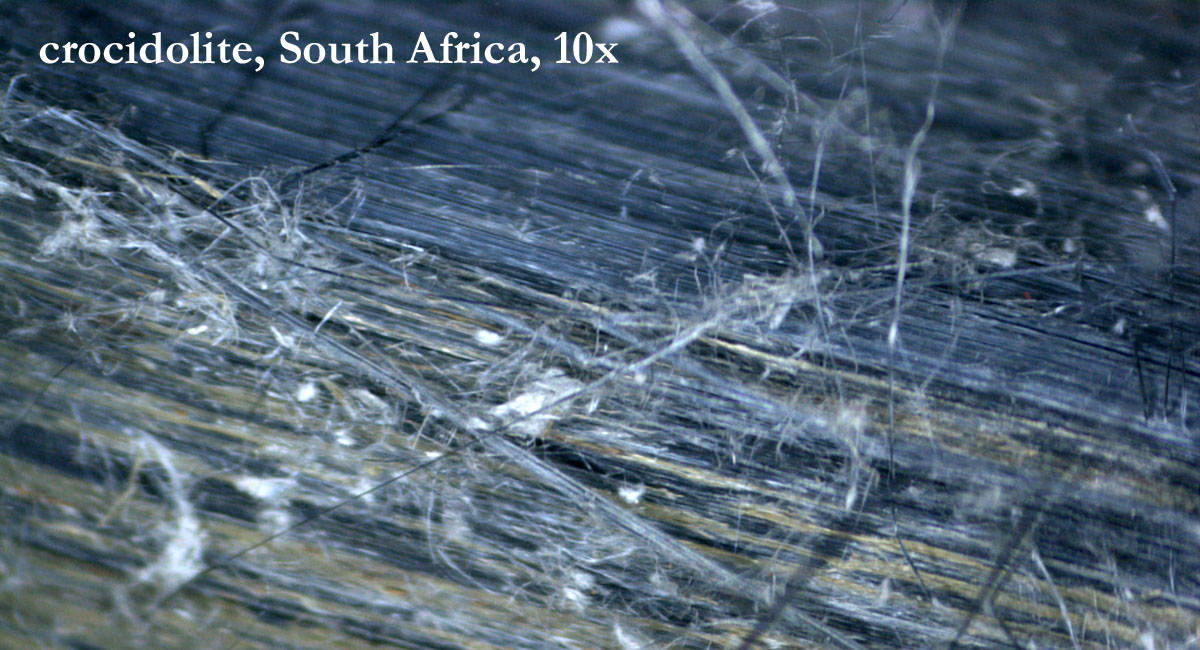
Chrysotile crocidolite amosite asbestos anthophyllite asbestos actinolite or. The asbestos materials are. When chrysotile asbestos is looked at under a microscope it wraps around itself in a spiral or serpentine shape.
This is the most common form of asbestos used commercially comprising about. In 120 cases material was prepared by alkali digestion and the residual dust was examined. Of inhaled chrysotile asbestos fibre.
Its chemical formula is mg 3 si 2 o 5oh 4 with some fe2 substituting for mg. The amount of iron substitution affects the refractive indices and the birefringence. The microscopy of asbestos.
Asbestos tremolite fibres under scanning electron microscopy.
More From Dora Coloring Pages Free Printable
- Cat Coloring Pages
- Mesothelioma Lawyer Michigan
- Uspto Attorney Lookup
- Mesothelioma Incurable
- Printable Turtle Coloring Sheet
Incoming Search Terms:
- Crocidolite Asbestos Printable Turtle Coloring Sheet,
- Chrysotile Wikipedia Printable Turtle Coloring Sheet,
- Asbestos Under The Microscope Printable Turtle Coloring Sheet,
- Analysis Of Fibres Using Microscopy Sciencedirect Printable Turtle Coloring Sheet,
- Chrysotile Asbestos Under A Microscope Imgur Printable Turtle Coloring Sheet,
- Anthophyllite An Overview Sciencedirect Topics Printable Turtle Coloring Sheet,
