Central Areas Of Whorled Collagen Fibers Mesothelioma, Http Tpx Sagepub Com Cgi Reprint 35 1 136 Pdf
Central areas of whorled collagen fibers mesothelioma Indeed lately has been sought by users around us, perhaps one of you. Individuals are now accustomed to using the net in gadgets to view image and video data for inspiration, and according to the title of this post I will talk about about Central Areas Of Whorled Collagen Fibers Mesothelioma.
- F The Disease Is Triggered By Environmental Antigens Powerpoint Presentation Free Online Download Ppt Zt1wfx
- Pneumoconiosis Ppt Download
- Mesothelioma Springerlink
- Respiratory Diseases
- Pneumoconiosis Springerlink
- 2
Find, Read, And Discover Central Areas Of Whorled Collagen Fibers Mesothelioma, Such Us:
- Pneumoconiosis Ppt Download
- Occupational Lung Disorder Authorstream
- Surgical Pathology Of Non Neoplastic Conditions Of The Pleura Pericardium And Peritoneum Chapter 6 Practical Pathology Of Serous Membranes
- Pneumoconiosis Ppt Download
- Pdf Mesothelioma In Domestic Animals Cytological And Anatomopathological Aspects
- Mesothelioma Va Pension
- Lawyer Dress For Kids
- Pleural Mesothelioma Lung
- Mens Costume Ideas
- Crown Coloring Page Printable
If you re searching for Crown Coloring Page Printable you've arrived at the perfect location. We have 100 images about crown coloring page printable adding images, photos, photographs, wallpapers, and more. In these webpage, we additionally have variety of graphics out there. Such as png, jpg, animated gifs, pic art, logo, black and white, translucent, etc.
A typical area in which the tumor cells are enmeshed in a network of thick collagen fibers.
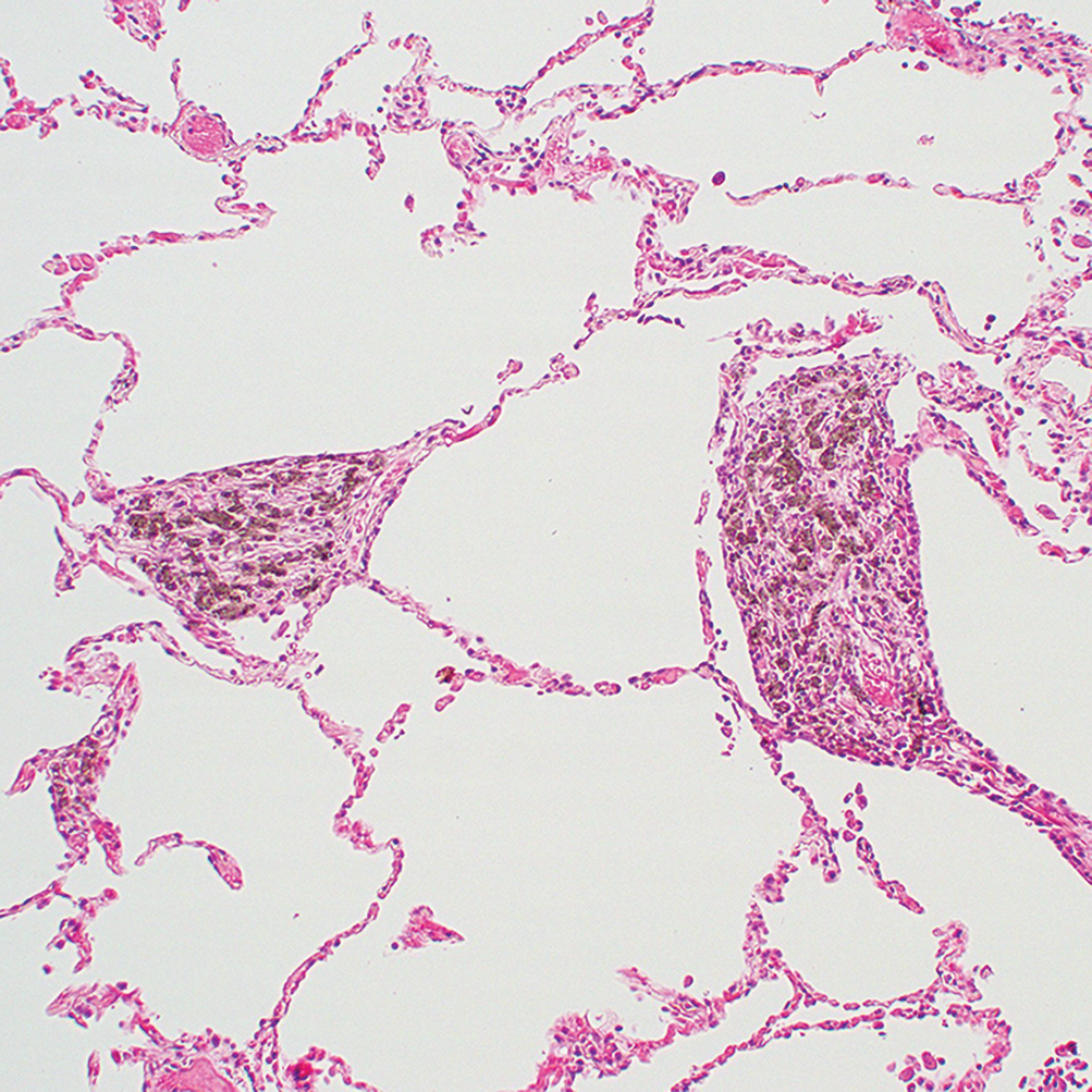
Crown coloring page printable. Central collagen becomes distinctly whorled and the relative number of inflammatory cells around the periphery decreases. In so called mixed dust pneumoconioses ie diseases caused. They were clearly positive in such a place as is shown in fig.
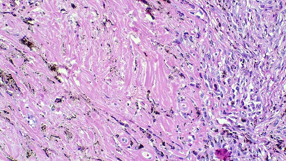
Central area of whorled collagen fibers with more peripheral zone of dust laden macrophages. 5 the thigh tumor shows a moderate cellularity with spindled nuclei in abundant collagenous stroma he 200. Asbestos bodies are encrusted with protein and iron.
Microscopically they consist of relatively acellular bundles of collagen in an undulating basketweave pattern and may contain abundant numbers of asbestos fibers almost exclusively chrysotile fibers but asbestos bodies are absent1 4 the inner side is covered by normal mesothelial cells and the costal side may demonstrate signs of. Fibers were surrounding the tumor cells inside the thick collagen fibers. Histochemical reactions mag showed type 1 predominance with more than 90 of type 1 fibers in 6 of 12 biopsy specimens.
Both alcian blue and colloid iron stains showed intercellular as well as in tracellular positivity. Whorled fibers as well as the fibers with a central loss of enzyme activity did not stain dark in the non specific esterase reaction. Building materials from older houses may contain asbestos which was used for years because of its fire retardant properties.
The positive material was completely. Whorls of reticulin and collagen fibers. 2 e f.
The central eosinophilic area of the rosettes is composed of collagen fibers he 100. 3 but less distinct in other areas. An example of a fibrous peritoneal mesothelioma having a benign histological appearance but nevertheless producing metastases is reported.
Central area of whorled collagen. The ferruginous bodies are asbestos fibers encrusted with iron. Microscopic morphology of silicosis.
Microscopy central area of whorled collagen fibers with a more peripheral zone of dust laden macrophages. The type 2 fiber loss could be confirmed using an antibody against serca1 fig. These ferruginous bodies are golden brown and beaded with a central colorless nonbirefringent core fiber.
Free radicals generated by asbestos fibers which preferentially localize in the distal lung close to the mesothelial layers. The dome of the diaphragm is covered by a pearly white. More peripheral zone of dust laden macrophages.
Golden brown fusiformbeaded robs with translucent center consisting of asbestos fibers coated with iron containing proteinaceous material.
More From Crown Coloring Page Printable
- Garbage Truck Coloring Page Free
- Princess Pinkie Pie Coloring Pages
- Sharma Law Firm
- Generation Of Reactive Oxygen Species By Human Mesothelioma Cells
- Eviction Lawyers Near Me
Incoming Search Terms:
- Pulmonary Pathology Flashcards Quizlet Eviction Lawyers Near Me,
- Https Academic Oup Com Ajcp Article Pdf 110 2 191 24883576 Ajcpath110 0191 Pdf Eviction Lawyers Near Me,
- Diffuse Interstitial Infiltrative Restrictive Diseases Pathology Eviction Lawyers Near Me,
- Pulmonary Pathology Flashcards Quizlet Eviction Lawyers Near Me,
- Http Doctor2015 Jumedicine Com Wp Content Uploads Sites 5 2018 01 Respiratory 17 Handout 5 Pdf Eviction Lawyers Near Me,
- Pulmonary Pathology Flashcards Quizlet Eviction Lawyers Near Me,

